Arrested pneumatization of the sphenoid sinus mimicking intraosseous lesions of the skull base
- Affiliations
-
- 1Department of Oral and Maxillofacial Radiology, University of Connecticut School of Dental Medicine, Farmington, CT, USA. tadinada@uchc.edu
- KMID: 2116794
- DOI: http://doi.org/10.5624/isd.2015.45.1.67
Abstract
- Arrested pneumatization of the sphenoid sinus is a developmental variant that is not always well recognized and is often confused with other pathologies associated with the skull base. This report describes the case of a patient referred for cone-beam computed tomography (CBCT) imaging for dental implant therapy. CBCT demonstrated a well-defined incidental lesion in the left sphenoid sinus with soft tissue-like density and sclerotic borders with internal curvilinear opacifications. The differential diagnoses included intraosseous lipoma, arrested pneumatization of the sphenoid sinus, chondrosarcoma, chondroid chordoma, and ossifying fibroma. The radiographic diagnosis of arrested pneumatization was based on the location of the lesion, its well-defined nature, the presence of internal opacifications, and lack of expansion. Gray-scale CBCT imaging of the area demonstrated values similar to fatty tissue. This case highlighted the fact that benign developmental variants associated with the skull base share similar radiographic features with more serious pathological entities.
MeSH Terms
Figure
-
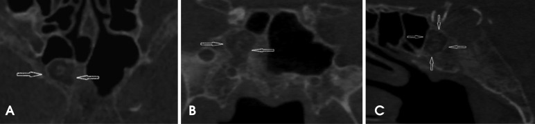
Fig. 1 The axial (A), coronal (B), and sagittal (C) images of a CBCT scan show the area of the arrested pneumatization of the right sphenoid sinus. The well-defined sclerotic borders, curvilinear internal calcifications, soft tissue density zones, and absence of any evidence of expansion or effect on the surrounding structures are visualized. The external morphology of the sphenoid bone appears to be normal.
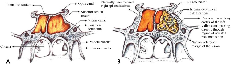
Fig. 2 A. A schematic diagram shows the normal pneumatization of the sphenoid sinus and the surrounding anatomic structures in this area. B. A schematic diagram shows arrested pneumatization of the left sphenoid sinus. The lesion has multiple foci of fat, narrow sclerotic margins, and internal curvilinear calcifications. The left vidian canal passes through the region of arrested pneumatization. Note the well-preserved bony cortex of the canal.
Reference
-
1. Spaeth J, Krugelstein U, Schlondorff G. The paranasal sinuses in CT-imaging: development from birth to age 25. Int J Pediatr Otorhinolaryngol. 1997; 39:25–40. PMID: 9051437.
Article2. Shah RK, Dhingra JK, Carter BL, Rebeiz EE. Paranasal sinus development: a radiographic study. Laryngoscope. 2003; 113:205–209. PMID: 12567069.
Article3. Scuderi AJ, Harnsberger HR, Boyer RS. Pneumatization of the paranasal sinuses: normal features of importance to the accurate interpretation of CT scans and MR images. AJR Am J Roentgenol. 1993; 160:1101–1104. PMID: 8470585.
Article4. Aoki S, Dillon WP, Barkovich AJ, Norman D. Marrow conversion before pneumatization of the sphenoid sinus: assessment with MR imaging. Radiology. 1989; 172:373–375. PMID: 2748818.
Article5. Jang YJ, Kim SC. Pneumatization of the sphenoid sinus in children evaluated by magnetic resonance imaging. Am J Rhinol. 2000; 14:181–185. PMID: 10887625.
Article6. Szolar D, Preidler K, Ranner G, Braun H, Kern R, Wolf G, et al. Magnetic resonance assessment of age-related development of the sphenoid sinus. Br J Radiol. 1994; 67:431–435. PMID: 8193887.
Article7. Degirmenci B, Haktanir A, Acar M, Albayrak R, Yücel A. Agenesis of sphenoid sinus: three cases. Surg Radiol Anat. 2005; 27:351–353. PMID: 16200385.
Article8. Welker KM, DeLone DR, Lane JI, Gilbertson JR. Arrested pneumatization of the skull base: imaging characteristics. AJR Am J Roentgenol. 2008; 190:1691–1696. PMID: 18492926.
Article9. Srubiski A, Csillag A, Timperley D, Kalish L, Qiu MR, Harvey RJ. Radiological features of the intraosseous lipoma of the sphenoid. Otolaryngol Head Neck Surg. 2011; 144:617–622. PMID: 21493245.
Article10. Politi M, Romeike BF, Papanagiotou P, Nabhan A, Struffert T, Feiden W, et al. Intraosseous hemangioma of the skull with dural tail sign: radiologic features with pathologic correlation. AJNR Am J Neuroradiol. 2005; 26:2049–2052. PMID: 16155158.11. Mah P, Reeves TE, McDavid WD. Deriving Hounsfield units using grey levels in cone beam computed tomography. Dentomaxillofac Radiol. 2010; 39:323–335. PMID: 20729181.
Article12. Reeves TE, Mah P, McDavid WD. Deriving Hounsfield units using grey levels in cone beam CT: a clinical application. Dentomaxillofac Radiol. 2012; 41:500–508. PMID: 22752324.
Article13. Taccone A, Oddone M, Occhi M, Dell'Acqua A, Ciccone MA. MRI "road-map" of normal age-related bone marrow. I. Cranial bone and spine. Pediatr Radiol. 1995; 25:588–595. PMID: 8570311.14. Kuntzler S, Jankowski R. Arrested pneumatization: witness of paranasal sinuses development? Eur Ann Otorhinolaryngol Head Neck Dis. 2014; 131:167–170. PMID: 24709406.
Article15. Yonetsu K, Watanabe M, Nakamura T. Age-related expansion and reduction in aeration of the sphenoid sinus: volume assessment by helical CT scanning. AJNR Am J Neuroradiol. 2000; 21:179–182. PMID: 10669247.16. Gurevitch O, Slavin S, Feldman AG. Conversion of red bone marrow into yellow - Cause and mechanisms. Med Hypotheses. 2007; 69:531–536. PMID: 17433565.
Article17. Buri´c N, Krasi´c D, Visnji´c M, Kati´c V. Intraosseous mandibular lipoma: a case report and review of the literature. J Oral Maxillofac Surg. 2001; 59:1367–1371. PMID: 11688046.18. Erdem E, Angtuaco EC, Van Hemert R, Park JS, Al-Mefty O. Comprehensive review of intracranial chordoma. Radiographics. 2003; 23:995–1009. PMID: 12853676.
Article19. Neff B, Sataloff RT, Storey L, Hawkshaw M, Spiegel JR. Chondrosarcoma of the skull base. Laryngoscope. 2002; 112:134–139. PMID: 11802051.
Article20. Daffner RH, Kirks DR, Gehweiler JA Jr, Heaston DK. Computed tomography of fibrous dysplasia. AJR Am J Roentgenol. 1982; 139:943–948. PMID: 6981980.
Article21. Politi M, Romeike BF, Papanagiotou P, Nabhan A, Struffert T, Feiden W, et al. Intraosseous hemangioma of the skull with dural tail sign: radiologic features with pathologic correlation. AJNR Am J Neuroradiol. 2005; 26:2049–2052. PMID: 16155158.22. Baumann I, Zimmermann R, Dammann F, Maassen MM. Ossifying fibroma of the ethmoid involving the orbit and the skull base. Otolaryngol Head Neck Surg. 2005; 133:158–159. PMID: 16025072.
Article23. Greenberg HS, Deck MD, Vikram B, Chu FC, Posner JB. Metastasis to the base of the skull: clinical findings in 43 patients. Neurology. 1981; 31:530–537. PMID: 6972014.24. Kösling S, Neumann K, Brandt S. CT and MRI of intrinsic space-occupying lesions of the bony skull base. Radiologe. 2009; 49:598–607. PMID: 19436984.
- Full Text Links
-
- Actions
-
Cited
- CITED
-
- Close
- Share
-
- Similar articles
-
- Arrested Pneumatization of the Sphenoid Sinus in the Skull Base
- A Case of Massive Skull Base Erosion by Fungus Ball in the Sphenoid Sinus
- Endoscopic Diagnosis and Treatment of Isolated Sphenoid Sinus Lesions
- A Case of Endoscopic Reconstruction of Skull Base Defect Combined with Meningoencephalocele in the Sphenoid Sinus
- Chondroma of the Sphenoid Sinus: Case Report
