Imaging Sci Dent.
2015 Mar;45(1):23-29. 10.5624/isd.2015.45.1.23.
Accuracy of virtual models in the assessment of maxillary defects
- Affiliations
-
- 1Department of Dentomaxillofacial Radiology, Faculty of Dentistry, Ankara University, Ankara, Turkey. dtkivo@yahoo.com
- 2Division of Dentomaxillofacial Radiology, Ministry of Health, Oral and Dental Health Center, Bolu, Turkey.
- 3Department of Anatomy, Gulhane Military Medical Academy, Ankara, Turkey.
- 4Department of Dentomaxillofacial Radiology, Dental Science Center, Gulhane Military Medical Academy, Ankara, Turkey.
- KMID: 2116788
- DOI: http://doi.org/10.5624/isd.2015.45.1.23
Abstract
- PURPOSE
This study aimed to assess the reliability of measurements performed on three-dimensional (3D) virtual models of maxillary defects obtained using cone-beam computed tomography (CBCT) and 3D optical scanning.
MATERIALS AND METHODS
Mechanical cavities simulating maxillary defects were prepared on the hard palate of nine cadavers. Images were obtained using a CBCT unit at three different fields-of-views (FOVs) and voxel sizes: 1) 60x60 mm FOV, 0.125 mm3 (FOV60); 2) 80x80 mm FOV, 0.160 mm3 (FOV80); and 3) 100x100 mm FOV, 0.250 mm3 (FOV100). Superimposition of the images was performed using software called VRMesh Design. Automated volume measurements were conducted, and differences between surfaces were demonstrated. Silicon impressions obtained from the defects were also scanned with a 3D optical scanner. Virtual models obtained using VRMesh Design were compared with impressions obtained by scanning silicon models. Gold standard volumes of the impression models were then compared with CBCT and 3D scanner measurements. Further, the general linear model was used, and the significance was set to p=0.05.
RESULTS
A comparison of the results obtained by the observers and methods revealed the p values to be smaller than 0.05, suggesting that the measurement variations were caused by both methods and observers along with the different cadaver specimens used. Further, the 3D scanner measurements were closer to the gold standard measurements when compared to the CBCT measurements.
CONCLUSION
In the assessment of artificially created maxillary defects, the 3D scanner measurements were more accurate than the CBCT measurements.
Keyword
MeSH Terms
Figure
-
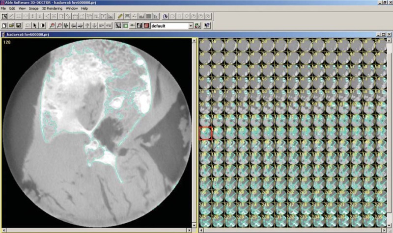
Fig. 1 Segmentation process with 3D Doctor
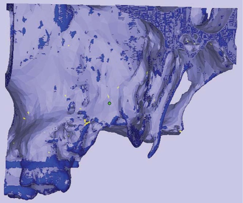
Fig. 2 Alignment of standardized field of view (FOV) images
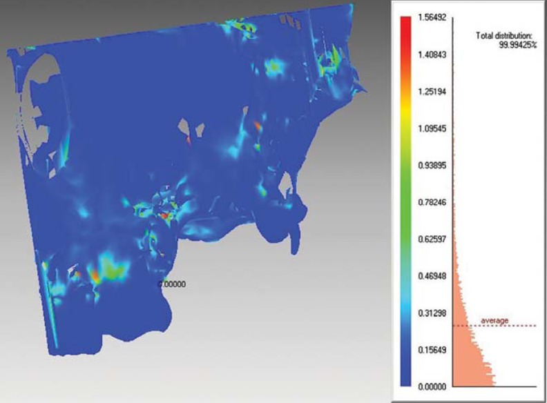
Fig. 3 Measurement of differences using standardized FOV images
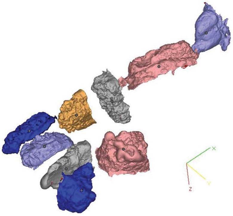
Fig. 4 Virtual models obtained from nine maxillary defects
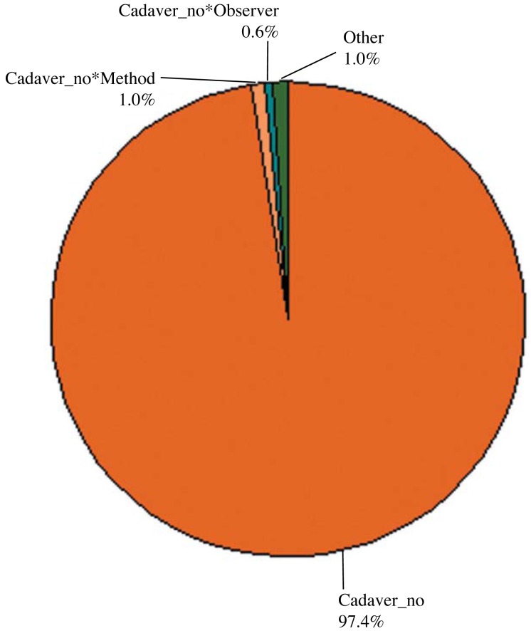
Fig. 5 Comparisons of different FOVs on the basis of observers
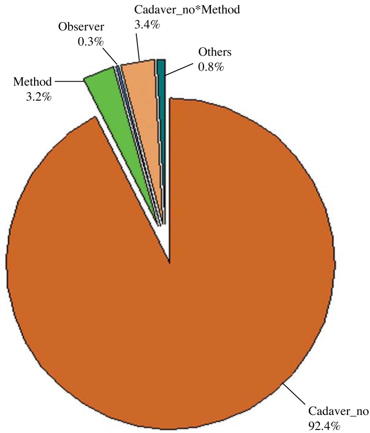
Fig. 6 Comparison of cone-beam computed tomography and three-dimensional scanner methods on the basis of observers and the gold standard
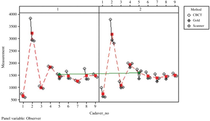
Fig. 7 Multi-vari chart for measurement by method/observer
Reference
-
1. Yuzbasıoglu E, Kurt H, Turunc R, Bilir H. Comparison of digital and conventional impression techniques: evaluation of patients' perception, treatment comfort, effectiveness and clinical outcomes. BMC Oral Health. 2014; 14:10. PMID: 24479892.
Article2. Lethaus B, Kessler P, Boeckman R, Poort LJ, Tolba R. Reconstruction of a maxillary defect with a fibula graft and titanium mesh using CAD/CAM techniques. Head Face Med. 2010; 6:16. PMID: 20642821.
Article3. Angelopoulos C, Scarfe WC, Farman AG. A comparison of maxillofacial CBCT and medical CT. Atlas Oral Maxillofac Surg Clin North Am. 2012; 20:1–17. PMID: 22365427.
Article4. Scarfe WC, Farman AG, Levin MD, Gane D. Essentials of maxillofacial cone beam computed tomography. Alpha Omegan. 2010; 103:62–67. PMID: 20645632.
Article5. Scarfe WC, Farman AG. What is cone-beam CT and how does it work? Dent Clin North Am. 2008; 52:707–730. PMID: 18805225.
Article6. Scarfe WC, Li Z, Aboelmaaty W, Scott SA, Farman AG. Maxillofacial cone beam computed tomography: essence, elements and steps to interpretation. Aust Dent J. 2012; 57(Suppl 1):46–60. PMID: 22376097.
Article7. Kamegawa M, Nakamura M, Fukui Y, Tsutsumi S, Hojo M. Direct 3-D morphological measurements of silicone rubber impression using micro-focus X-ray CT. Dent Mater J. 2010; 29:68–74. PMID: 20379015.
Article8. Boldt F, Weinzierl C, Hertrich K, Hirschfelder U. Comparison of the spatial landmark scatter of various 3D digitalization methods. J Orofac Orthop. 2009; 70:247–263. PMID: 19484417.
Article9. Barone S, Paoli A, Razionale AV. Creation of 3D multi-body orthodontic models by using independent imaging sensors. Sensors (Basel). 2013; 13:2033–2050. PMID: 23385416.
Article10. Motohashi N, Kuroda T. A 3D computer-aided design system applied to diagnosis and treatment planning in orthodontics and orthognathic surgery. Eur J Orthod. 1999; 21:263–274. PMID: 10407535.
Article11. Lu P, Li Z, Wang Y, Chen J, Zhao J. The research and development of noncontact 3-D laser dental model measuring and analyzing system. Chin J Dent Res. 2000; 3:7–14. PMID: 11314539.12. Hirogaki Y, Sohmura T, Satoh H, Takahashi J, Takada K. Complete 3-D reconstruction of dental cast shape using perceptual grouping. IEEE Trans Med Imaging. 2001; 20:1093–1101. PMID: 11686444.
Article13. Agbaje JO, Jacobs R, Michiels K, Abu-Ta'a M, van Steenberghe D. Bone healing after dental extractions in irradiated patients: a pilot study on a novel technique for volume assessment of healing tooth sockets. Clin Oral Investig. 2009; 13:257–261.
Article14. Turbush SK, Turkyilmaz I. Accuracy of three different types of stereolithographic surgical guide in implant placement: an in vitro study. J Prosthet Dent. 2012; 108:181–188. PMID: 22944314.
Article15. Pohlenz P, Blessmann M, Blake F, Gbara A, Schmelzle R, Heiland M. Major mandibular surgical procedures as an indication for intraoperative imaging. J Oral Maxillofac Surg. 2008; 66:324–329. PMID: 18201617.
Article16. Katsumata A, Hırukawa A, Okumura S, Naitoh M, Fujishita M, Ariji E, et al. Effects of image artifacts on gray-value density in limited-volume cone-beam computerized tomography. Oral Surg Oral Med Oral Pathol Oral Radiol Endod. 2007; 104:829–836. PMID: 17448704.
Article17. Kwong JC, Palomo JM, Landers MA, Figueroa A, Hans MG. Image quality produced by different cone-beam computed tomography settings. Am J Orthod Dentofacial Orthop. 2008; 133:317–327. PMID: 18249300.
Article18. Hassan B, Couto Souza P, Jacobs R, de Azambuja Berti S, van der Stelt P. Influence of scanning and reconstruction parameters on quality of three-dimensional surface models of the dental arches from cone beam computed tomography. Clin Oral Investig. 2010; 14:303–310.
Article19. Sezgin OS, Kayıpmaz S, Sahin B. The effect of slice thickness on the assessment of bone defect volumes by the Cavalieri principle using cone beam computed tomography. J Digit Imaging. 2013; 26:115–118. PMID: 22539100.
Article20. Emirzeoglu M, Sahin B, Selcuk MB, Kaplan S. The effects of section thickness on the estimation of liver volume by the Cavalieri principle using computed tomography images. Eur J Radiol. 2005; 56:391–397. PMID: 15893441.
Article21. Sahin B, Mazonakis M, Akan H, Kaplan S, Bek Y. Dependence of computed tomography volume measurements upon section thickness: an application to human dry skulls. Clin Anat. 2008; 21:479–485. PMID: 18627101.
Article22. Loubele M, Maes F, Schutyser F, Marchal G, Jacobs R, Suetens P. Assessment of bone segmentation quality of conebeam CT versus multislice spiral CT: a pilot study. Oral Surg Oral Med Oral Pathol Oral Radiol Endod. 2006; 102:225–234. PMID: 16876067.
Article23. Pinsky HM, Dyda S, Pinsky RW, Misch KA, Sarment DP. Accuracy of three-dimensional measurements using conebeam CT. Dentomaxillofac Radiol. 2006; 35:410–416. PMID: 17082331.
Article
- Full Text Links
-
- Actions
-
Cited
- CITED
-
- Close
- Share
-
- Similar articles
-
- An Evaluation of Validity of Measurements using Digital Caliper and Three-dimensional Virtual Dental Models
- Integrated three-dimensional digital assessment of accuracy of anterior tooth movement using clear aligners
- Accuracy of conventional and digital mounting of dental models: A literature review
- Trueness of 3D printed partial denture frameworks: build orientations and support structure density parameters
- Recording maximal intercuspation and border positions of the mandible with intraoral scanner using the acquisition software’s multi-occlusion function
