Ultrasound-Guided Intervention in Lumbar Spine
- Affiliations
-
- 1Department of Orthopaedic Surgery, Kwangju Christian Hospital, Gwangju, Korea. stemcellchoi@gmail.com
- KMID: 2106745
- DOI: http://doi.org/10.4055/jkoa.2015.50.2.107
Abstract
- For precise diagnosis and therapeutic interventions for chronic lumbar pain, lumbar nerve block has been performed primarily by computed tomography or C-arm fluoroscopy. The use of such equipment has disadvantages in that both patient and surgeon are exposed to radiation, and it could be used in limited areas. The current role of ultrasound guidance for musculoskeletal intervention treatments has been reported. Recently, to compensate for such disadvantage and to be able to approach easily in the outpatient department, interest in ultrasound-guided lumbar nerve block is increasing, and several studies have reported relatively high success rates. However, ultrasound-guided intervention for lumbar nerve block has not been exactly described. Therefore, we conducted this review to evaluate the feasibility of ultrasound guided lumbar interventions.
Keyword
Figure
-

Figure 1 Longitudinal facet views were obtained by curved transducer for indentification of the different spinal segments (A) and the longitudinal facet view shows L3-4, L4-5 and L5-S1 facet joint (B).
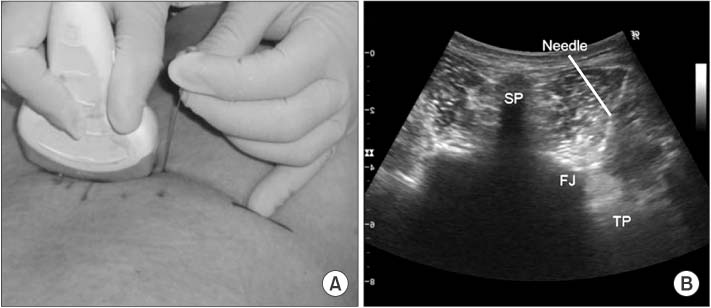
Figure 2 Needle insertion between the superior articular process and on the upper edge of the transverse process. (A) Procedure photo. (B) Ultrasound finding at needle insertion. SP, spinous process; FJ, facet joint; TP, transverse process.

Figure 3 The fluoroscopy showed correct insertion of the needle for medial branch block and distribution of the contrast media (0.5 ml).
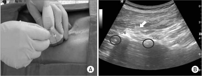
Figure 4 The needle for selective nerve root block was inserted at the same angle with the needle for medial branch block as short axis out of plane approach. (A) Procedure photo. (B) Ultrasound finding at needle insertion. TP, transverse process.

Figure 5 The position of the needles and distribution of the contrast medium was evaluated with C-arm fluoroscopy. The two arrows indicate needles for medial branch block and the two arrowheads for selective nerve root block.

Figure 6 The transducer was placed transversely on the sacral hiatus and checked intercornual distance, thickness of sacrococcygeal membrane, depth of caudal space. (A) Procedure photo. (B) Ultrasound finding.
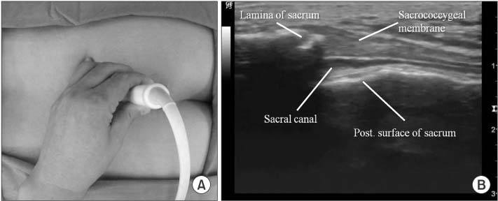
Figure 7 The transducer was rotated 90 degrees to obtain the longitudinal view of sacral hiatus. (A) Procedure photo. (B) Ultrasound finding.

Figure 8 A needle was inserted to the caudal epidural space under ultrasound guidance. (A) Procedure photo.(B) Ultrasound finding at needle insertion.
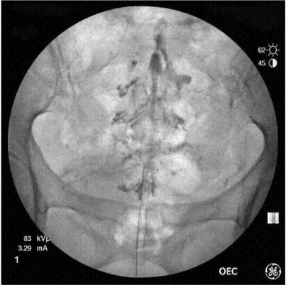
Figure 9 Contrast media was spread into the sacral canal and Christmas-tree like appearance was observed.
Reference
-
1. Peterson MK, Millar FA, Sheppard DG. Ultrasound-guided nerve blocks. Br J Anaesth. 2002; 88:621–624.2. Greher M, Kirchmair L, Enna B, et al. Ultrasound-guided lumbar facet nerve block: accuracy of a new technique confirmed by computed tomography. Anesthesiology. 2004; 101:1195–1200.3. Greher M, Scharbert G, Kamolz LP, et al. Ultrasound-guided lumbar facet nerve block: a sonoanatomic study of a new methodologic approach. Anesthesiology. 2004; 100:1242–1248.4. Ha DH, Shim DM, Kim TK, Kim YM, Choi SS. Comparison of ultrasonography- and fluoroscopy-guided facet joint block in the lumbar spine. Asian Spine J. 2010; 4:15–22.
Article5. Jung H, Jeon S, Ahn S, Kim M, Choi Y. The validation of ultrasound-guided lumbar facet nerve blocks as confirmed by fluoroscopy. Asian Spine J. 2012; 6:163–167.
Article6. Kim D, Choi D, Kim C, Kim J, Choi Y. Transverse process and needles of medial branch block to facet joint as landmarks for ultrasound-guided selective nerve root block. Clin Orthop Surg. 2013; 5:44–48.
Article7. Jung H, Kim DH, Jeon SH, Kim CY, Kim JS, Choi YS. The effectiveness of ultrasound guidance in caudal epidural block. J Korean Soc Spine Surg. 2013; 20:178–183.
Article8. Helbig T, Lee CK. The lumbar facet syndrome. Spine (Phila Pa 1976). 1988; 13:61–64.
Article9. Saal JS. General principles of diagnostic testing as related to painful lumbar spine disorders: a critical appraisal of current diagnostic techniques. Spine (Phila Pa 1976). 2002; 27:2538–2545.10. Sato M, Simizu S, Kadota R, Takahasi H. Ultrasound and nerve stimulation-guided L5 nerve root block. Spine (Phila Pa 1976). 2009; 34:2669–2673.
Article11. Riew KD, Park JB, Cho YS, et al. Nerve root blocks in the treatment of lumbar radicular pain. A minimum five-year follow-up. J Bone Joint Surg Am. 2006; 88:1722–1725.12. Gray AT. Ultrasound-guided regional anesthesia: current state of the art. Anesthesiology. 2006; 104:368–373.13. Marhofer P, Greher M, Kapral S. Ultrasound guidance in regional anaesthesia. Br J Anaesth. 2005; 94:7–17.14. White AH, Derby R, Wynne G. Epidural injections for the diagnosis and treatment of low-back pain. Spine (Phila Pa 1976). 1980; 5:78–86.
Article15. Weinstein SM, Herring SA, Derby R. Contemporary concepts in spine care. Epidural steroid injections. Spine (Phila Pa 1976). 1995; 20:1842–1846.16. Standring S. Gray's anatomy. The anatomical basis of clinical practice. 39th ed. New York: Churchill Livingstone;2005. p. 750.17. Stitz MY, Sommer HM. Accuracy of blind versus fluoroscopically guided caudal epidural injection. Spine (Phila Pa 1976). 1999; 24:1371–1376.
Article18. Lewis MP, Thomas P, Wilson LF, Mulholland RC. The 'whoosh' test A clinical test to confirm correct needle placement in caudal epidural injections. Anaesthesia. 1992; 47:57–58.19. Tsui BC, Tarkkila P, Gupta S, Kearney R. Confirmation of caudal needle placement using nerve stimulation. Anesthesiology. 1999; 91:374–378.
Article20. Riew KD, Yin Y, Gilula L, et al. The effect of nerve-root injections on the need for operative treatment of lumbar radicular pain. A prospective, randomized, controlled, double-blind study. J Bone Joint Surg Am. 2000; 82:1589–1593.21. Jarvik JG, Deyo RA. Diagnostic evaluation of low back pain with emphasis on imaging. Ann Intern Med. 2002; 137:586–597.
Article22. Fenton DS, Czervionke LF. Image-guided spine intervention. Philadelphia: Saunders;2003. p. 73–97.23. Nakagawa M, Shinbori H, Ohseto K. Ultrasound-guided and fluoroscopy-assisted selective cervical nerve root blocks. Masui. 2009; 58:1506–1511.24. Narouze SN, Vydyanathan A, Kapural L, Sessler DI, Mekhail N. Ultrasound-guided cervical selective nerve root block: a fluoroscopy-controlled feasibility study. Reg Anesth Pain Med. 2009; 34:343–348.25. Shim JK, Moon JC, Yoon KB, Kim WO, Yoon DM. Ultrasound-guided lumbar medial-branch block: a clinical study with fluoroscopy control. Reg Anesth Pain Med. 2006; 31:451–454.
Article26. Klocke R, Jenkinson T, Glew D. Sonographically guided caudal epidural steroid injections. J Ultrasound Med. 2003; 22:1229–1232.
Article27. Chen CP, Tang SF, Hsu TC, et al. Ultrasound guidance in caudal epidural needle placement. Anesthesiology. 2004; 101:181–184.
Article28. Roh JH, Kim WO, Yoon KB, Yoon DM. The success rate of caudal block under ultrasound guidance and the direction of the needle in the sacral canal. Korean J Pain. 2007; 20:40–45.
Article29. Roberts SA, Guruswamy V, Galvez I. Caudal injectate can be reliably imaged using portable ultrasound: a preliminary study. Paediatr Anaesth. 2005; 15:948–952.30. Pettit AC, Kropski JA, Castilho JL, et al. The index case for the fungal meningitis outbreak in the United States. N Engl J Med. 2012; 367:2119–2125.
Article31. Friedly J, Chan L, Deyo R. Increases in lumbosacral injections in the Medicare population: 1994 to 2001. Spine (Phila Pa 1976). 2007; 32:1754–1760.32. Shelokov AP. Re: Friedly J, Chan L, Deyo R. Increases in lumbosacral injections in the Medicare population: 1994 to 2001. Spine 2007;32:1754-60. Spine (Phila Pa 1976). 2007; 32:3090–3091.33. Weinstein JN, Tosteson TD, Lurie JD, et al. Surgical versus nonoperative treatment for lumbar spinal stenosis four-year results of the Spine Patient Outcomes Research Trial. Spine (Phila Pa 1976). 2010; 35:1329–1338.
Article34. Radcliff KE, Rihn J, Hilibrand A, et al. Does the duration of symptoms in patients with spinal stenosis and degenerative spondylolisthesis affect outcomes?: analysis of the Spine Outcomes Research Trial. Spine (Phila Pa 1976). 2011; 36:2197–2210.35. Dadure C, Raux O, Rochette A, Capdevila X. Interest of ultrasonographic guidance in paediatric regional anaesthesia. Ann Fr Anesth Reanim. 2009; 28:878–884.36. de Josemaría B, Gálvez I, Reinoso-Barbero F. Ultrasound guidance in pediatric regional anesthesia. Rev Esp Anestesiol Reanim. 2009; 56:170–179.37. Schwartz D, Raghunathan K, Dunn S, Connelly NR. Ultrasonography and pediatric caudals. Anesth Analg. 2008; 106:97–99.
Article
