Intramedullary Spinal Lesions Involving the Conus Medullaris: MR Imaging Features for Differential Diagnosis
- Affiliations
-
- 1Department of Radiology, Gangnam Severance Hospital, Yonsei University College of Medicine, Seoul, Korea. suhsh11@yuhs.ac
- 2Department of Neurosurgery, Gangnam Severance Hospital, Yonsei University, Seoul, Korea.
- 3Severance Institute of Vascular and Metabolic Research, Yonsei University College of Medicine, Seoul, Korea.
- KMID: 2099890
- DOI: http://doi.org/10.13104/jksmrm.2014.18.2.144
Abstract
- PURPOSE
Intramedullary spinal lesions in the conus medullaris (CM), including tumors and vascular lesion, are rarely reported. We reported various MR features of intramedullary spinal cord lesions involving the CM including ependymoma, hemangioblastomas, dermoid cyst, ventriculus terminalis and spinal AVF and tried to discuss them for differential diagnosis.
MATERIALS AND METHODS
Six patients (male: female = 4:2, mean age = 44.3 year old) were enrolled from the clinical database of our institute from 2004 to 2010 and their radiological images and clinical symptoms were reviewed retrospectively. All patients had taken initial and postoperative MRI with contrast enhancement using gadopentate dimeglumine (Gd-DTPA). These images were analyzed by tumor size, location, signal intensity relative to the spinal cord, vascular flow voids, syrinx or cyst, edema and enhancement pattern.
RESULTS
Contrast enhancement was seen in all intramedullary masses. An eccentric enhancing nodule was noted in two hemangioblastomas and unusual peripheral rim enhancement with septation was seen in ventriculus terminalis. Patchy enhancement of the CM was observed in spinal arteriovenous fistula (AVF). Extensive cord edema adjacent to the intramedullary lesions was seen in four cases and syrinx was noted in three cases. Vascular signal voids were found in two hemangioblastomas and one spinal AVF.
CONCLUSION
In evaluation of intramedullary spinal lesions in the CM, it is necessary to consider these unusual MR findings and discriminate various pathologies with prudence and caution.
Keyword
MeSH Terms
Figure
-
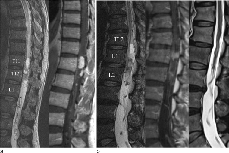
Fig. 1 Intramedullary hemangioblastoma (a) versus Spinal arteriovenous shunt (b). a. T2-weighted sagittal image (left) shows solid mass of the conus medullaris with hydrosyrinx, extensive cord edema and dilated medullary veins. Contrast enhanced T1-weighted sagittal image (right) shows enhancement of two solid components. b. T2-weighted sagittal image (left) shows swelling and high signal intensity and atrophy of the conus medullaris with hydrosyrinx, dilated medullary vein and proximal cord edema. Contrast enhanced T1-weighted sagittal image (middle) shows focal enhancement of the conus medullaris, probably due to chronic ischemic insult from venous hypertension. On follow-up MR (right) cord edema and syrinx was disappeared after embolization.

Fig. 2 Ventriculus terminalis (a) versus Intramedullary hemangioblastoma (b). a. T2-weighted sagittal image (left) shows cystic mass in the conus medullaris without hydrosyrinx and cord edema. Contrast enhanced T1-weighted sagittal image (right) shows peripheral rim enhancement of the cyst with internal septum. b. T2-weighted sagittal image (left) shows cystic mass of the conus medullaris with extensive cord edema and dilated perimedullary vein. Contrast enhanced T1-weighted sagittal image (right) shows eccentric enhancement of the nodular component within the intratumoral cyst.

Fig. 3 Myxopapillary ependymoma (a) versus Dermoid cyst (b). a. T2-weighted sagittal image (left) shows solid and cystic mass in the conus medullaris with extensive hydrosyrinx and cord edema. Contrast enhanced T1-weighted sagittal image (right) shows enhancement of the solid component. b. T1-weighted sagittal image (left) shows heterogeneous signal intensity within the intratumoral cyst and T2-weighted sagittal image (middle) shows solid and cystic mass of the conus medullaris without hydrosyrinx. Contrast enhanced T1-weighted sagittal image (right) shows rim enhancement of the solid component.
Reference
-
1. Admiraal P, Hazenberg GJ, Algra PR, Kamphorst W, Wolbers JG. Spinal subarachnoid hemorrhage due to a filum terminale ependymoma. Clin Neurol Neurosurg. 1992; 94:69–72.2. Hassan MF, Mohamed MB, Kalsi P, Sinar EJ, Bradey N. Intramedullary pyogenic abscess in the conus medullaris. Br J Neurosurg. 2012; 26:118–119.3. Sharma M, Mally R, Velho V. Ruptured conus medullaris dermoid cyst with fat droplets in the central canal. Asian Spine J. 2013; 7:50–54.4. Chen CY, Chen PH, Yao MS, Chu JS, Chan WP. MRI of hemangioblastoma in the conus medullaris. Comput Med Imaging Graph. 2008; 32:78–81.5. Welling LC, Zanellato C, Tessari M, Mendes V, Figueiredo EG, Teixeira MJ. Hemangioblastoma of the conus medullaris. Br J Neurosurg. 2012; 26:296–297.6. Shimosawa H, Matsumoto M, Yabe H, Mukai M, Toyama Y, Morioka H. Primary primitive neuroectodermal tumor of the conus medullaris in an elderly patient: a case report and review of the literature. Case Rep Oncol. 2011; 4:267–274.7. Sanborn MR, Pramick M, Brooks J, Welch WC. Glioblastoma multiforme in the adult conus medullaris. J Clin Neurosci. 2011; 18:842–843.8. Jaiswal AK, Jaiswal S, Gupta SK, Singh Gautam VK, Kumar S. Intramedullary tuberculoma of the conus. J Clin Neurosci. 2006; 13:870–872.9. Kahilogullari G, Erdem A, Heper AO, Erden E. Intramedullary mature cystic teratoma of the conus medullaris. A case report. J Neurosurg Sci. 2006; 50:55–58.10. Gallia GL, Burger PC, Suk I, et al. Concomitant conus medullaris ependymoma and filum terminale lipoma: case report. Neurosurgery. 2006; 58:E1214.11. Srivatanakul K, Songsaeng D, Ozanne A, Toulgoat F, Lasjaunias P. Spinal arteriovenous malformation associated with syringomyelia. J Neurosurg Spine. 2009; 10:436–442.12. Oh MC, Kim JM, Kaur G, et al. Prognosis by tumor location in adults with spinal ependymomas. J Neurosurg Spine. 2013; 18:226–235.13. Kim DH, Kim JH, Choi SH, et al. Differentiation between intramedullary spinal ependymoma and astrocytoma: comparative MRI analysis. Clin Radiol. 2014; 69:29–35.14. Goy AM, Pinto RS, Raghavendra BN, Epstein FJ, Kricheff II. Intramedullary spinal cord tumors: MR imaging, with emphasis on associated cysts. Radiology. 1986; 161:381–386.15. Sun B, Wang C, Wang J, Liu A. MRI features of intramedullary spinal cord ependymomas. J Neuroimaging. 2003; 13:346–351.16. Koeller KK, Rosenblum RS, Morrison AL. Neoplasms of the spinal cord and filum terminale: radiologic-pathologic correlation. Radiographics. 2000; 20:1721–1749.17. Epstein FJ, Farmer JP, Freed D. Adult intramedullary spinal cord ependymomas: the result of surgery in 38 patients. J Neurosurg. 1993; 79:204–209.18. Kahan H, Sklar EM, Post MJ, Bruce JH. MR characteristics of histopathologic subtypes of spinal ependymoma. AJNR Am J Neuroradiol. 1996; 17:143–150.19. Browne TR, Adams RD, Roberson GH. Hemangioblastoma of the spinal cord. Review and report of five cases. Arch Neurol. 1976; 33:435–441.20. Baker KB, Moran CJ, Wippold FJ 2nd, et al. MR imaging of spinal hemangioblastoma. AJR Am J Roentgenol. 2000; 174:377–382.21. Park CH, Lee CH, Hyun SJ, Jahng TA, Kim HJ, Kim KJ. Surgical outcome of spinal cord hemangioblastomas. J Korean Neurosurg Soc. 2012; 52:221–227.22. Murota T, Symon L. Surgical management of hemangioblastoma of the spinal cord: a report of 18 cases. Neurosurgery. 1989; 25:699–707.23. Fine MJ, Kricheff II, Freed D, Epstein FJ. Spinal cord ependymomas: MR imaging features. Radiology. 1995; 197:655–658.24. Yoshii S, Shimizu K, Ido K, Nakamura T. Ependymoma of the spinal cord and the cauda equina region. J Spinal Disord. 1999; 12:157–161.25. Han IH, Kuh SU, Chin DK, Kim KS, Jin BH, Cho YE. Surgical treatment of primary spinal tumors in the conus medullaris. J Korean Neurosurg Soc. 2008; 44:72–77.26. Lowe GM. Magnetic resonance imaging of intramedullary spinal cord tumors. J Neurooncol. 2000; 47:195–210.27. Bostrom A, Hans FJ, Reinacher PC, et al. Intramedullary hemangioblastomas: timing of surgery, microsurgical technique and follow-up in 23 patients. Eur Spine J. 2008; 17:882–886.28. Xu QW, Bao WM, Mao RL, Yang GY. Magnetic resonance imaging and microsurgical treatment of intramedullary hemangioblastoma of the spinal cord. Neurosurgery. 1994; 35:671–675.29. Samii M, Klekamp J. Surgical results of 100 intramedullary tumors in relation to accompanying syringomyelia. Neurosurgery. 1994; 35:865–873.30. Brisman JL, Borges LF, Ogilvy CS. Extramedullary hemangioblastoma of the conus medullaris. Acta Neurochir (Wien). 2000; 142:1059–1062.31. Sigal R, Denys A, Halimi P, Shapeero L, Doyon D, Boudghene F. Ventriculus terminalis of the conus medullaris: MR imaging in four patients with congenital dilatation. AJNR Am J Neuroradiol. 1991; 12:733–737.32. Matsubayashi R, Uchino A, Kato A, Kudo S, Sakai S, Murata S. Cystic dilatation of ventriculus terminalis in adults: MRI. Neuroradiology. 1998; 40:45–47.33. Dullerud R, Server A, Berg-Johnsen J. MR imaging of ventriculus terminalis of the conus medullaris. A report of two operated patients and a review of the literature. Acta Radiol. 2003; 44:444–446.34. Liccardo G, Ruggeri F, De Cerchio L, Floris R, Lunardi P. Fifth ventricle: an unusual cystic lesion of the conus medullaris. Spinal Cord. 2005; 43:381–384.35. Brisman JL, Li M, Hamilton D, Mayberg MR, Newell DW. Cystic dilation of the conus ventriculus terminalis presenting as an acute cauda equina syndrome relieved by decompression and cyst drainage: case report. Neurosurgery. 2006; 58:E585.36. Sansur CA, Sheehan JP, Sherman JH, Jane JA. Ventriculus terminalis causing back pain and urinary retention. Acta Neurochir (Wien). 2006; 148:919–920.37. Ciappetta P, D'Urso PI, Luzzi S, Ingravallo G, Cimmino A, Resta L. Cystic dilation of the ventriculus terminalis in adults. J Neurosurg Spine. 2008; 8:92–99.38. Dhillon RS, McKelvie PA, Wang YY, Han T, Murphy M. Cystic lesion of the ventriculus terminalis in an adult. J Clin Neurosci. 2010; 17:1601–1603.39. van Aalst J, Hoekstra F, Beuls EA, et al. Intraspinal dermoid and epidermoid tumors: report of 18 cases and reappraisal of the literature. Pediatr Neurosurg. 2009; 45:281–290.40. Graham DV, Tampieri D, Villemure JG. Intramedullary dermoid tumor diagnosed with the assistance of magnetic resonance imaging. Neurosurgery. 1988; 23:765–767.41. Muthukumar N, Srisaravanan J. Intramedullary dermoid in a low lying conus tethered by a fatty filum - embryological implications. Acta Neurochir (Wien). 2007; 149:1173–1175.42. Krishna KK, Agarwal PA, Agarwal SI, Jain MM. Dermoid of the conus medullaris. J Clin Neurosci. 2004; 11:796–797.43. Patankar AP, Sheth JH. Dermoid cyst: a rare intramedullary inclusion cyst. Asian J Neurosurg. 2012; 7:81–83.
- Full Text Links
-
- Actions
-
Cited
- CITED
-
- Close
- Share
-
- Similar articles
-
- Intramedullary Spinal Cystic Teratoma of the Conus Mudullaris with Caudal Exophytic Growth: Case Report
- Intramedullary Neurilemmoma in Conus Medullaris
- An Intramedullary Neurenteric Cyst in the Conus Medullaris with Recurrent Meningitis
- Ganglioglioma of Conus Medullaris: Case Report
- Multiple Intramedullary and Intradural Epidermoid Cysts in the Conus Medullaris and the Lumbar Spine: Case Report
