Calcific Tendinitis of the Hand and Foot: A Report of Four Cases
- Affiliations
-
- 1Department of Radiology, School of Medicine, Catholic University of Daegu, Korea. yhlee@cu.ac.kr
- 2Department of Radiology, Pohang St. Mary's Hospital, Korea.
- 3Department of Radiology, School of Medicine, Dongguk University Gyeongju Hospital, Korea.
- KMID: 2099853
- DOI: http://doi.org/10.13104/jksmrm.2012.16.2.177
Abstract
- Calcific tendinitis of hand and foot is rare and frequently misdiagnosed because of its rare incidence and its similar clinical presentation to other conditions such as infection. Awareness of the typical location as well as familiarity with the imaging findings is essential for making a correct diagnosis of this rare condition. We report four cases of calcific tendinitis of hand and foot, occurring in the flexor hallucis brevis, abductor digiti minimi, and abductor pollicis brevis.
Keyword
Figure
-
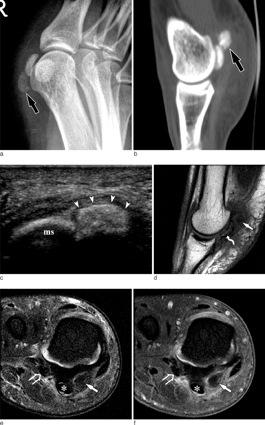
Fig. 1 A 32-year-old woman with calcific tendinitis of the medial head of flexor hallucis brevis. Oblique radiograph (a) and CT with sagittal reconstruction (b) of the right first metatarsophalangeal joint show an amorphous calcification (arrow) adjacent to the medial sesamoid bone. c. US reveals an ovoid hyperechoic calcification (arrowheads) within the medial head of flexor hallucis brevis tendon. d. On sagittal T1-weighted MR image, calcification is seen as a faint low signal intensity nodule. Soft tissue edema is seen around the calcification and bone marrow edema is also seen in the medial sesamoid bone (curved arrow). Coronal fat-suppressed T2-weighted (e) and contrast-enhanced fat-suppressed T1-weighted (f) images show diffuse soft tissue edema and enhancement around the calcification (arrow) in the medial head of flexor hallucis brevis tendon. The flexor hallucis longus (asterisk) and lateral head of flexor hallucis brevis (open arrow) tendons are indicated. ms = medial sesamoid.
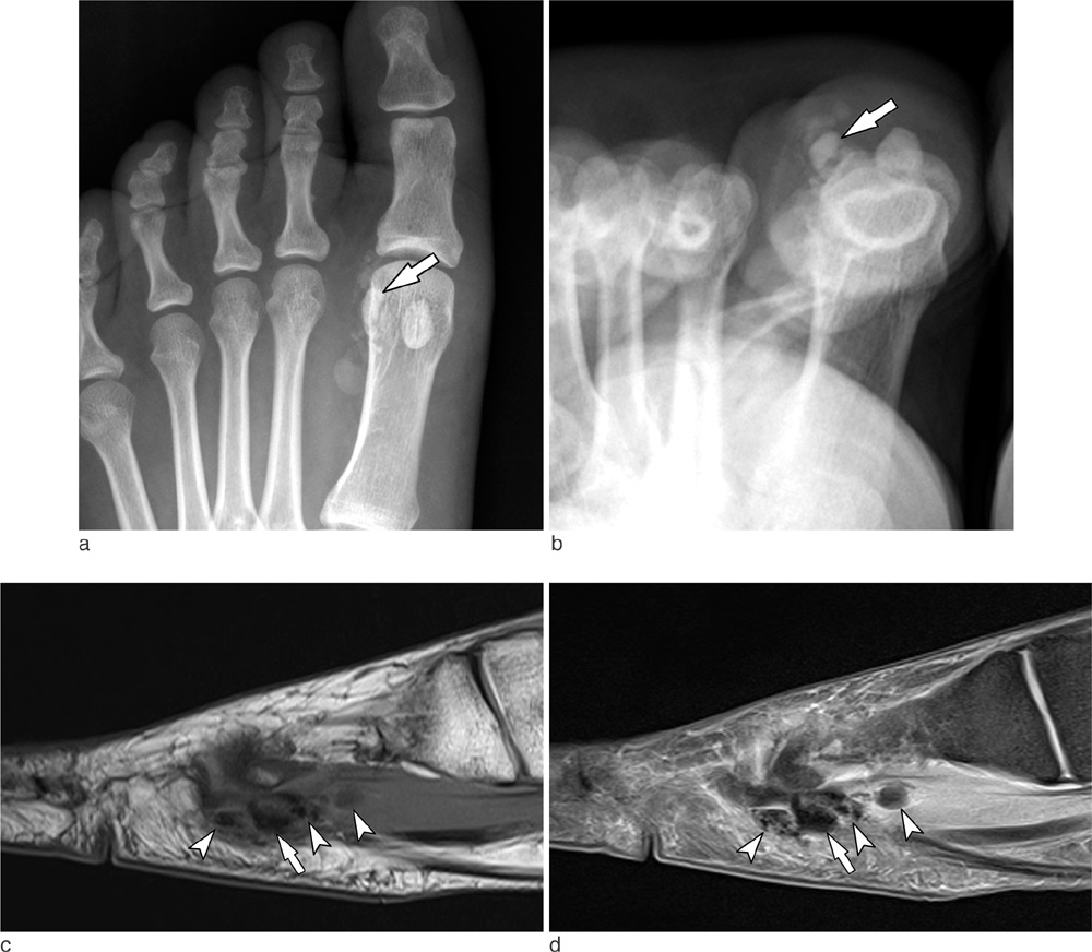
Fig. 2 A 42-year-old woman with calcific tendinitis of the lateral head of flexor hallucis brevis. a, b. Anteroposterior and tangential radiographs of the left first metatarsophalangeal joint show stippled and ovoid calcifications around the lateral sesamoid bone (arrow). Sagittal T1-weighted (c) and fat-suppressed proton density-weighted (d) images show irregular calcifications (arrowheads) in the lateral head of the flexor hallucis brevis tendon and muscle and soft tissue edema around the calcifications. The lateral sesamoid bone is indicated (arrow).
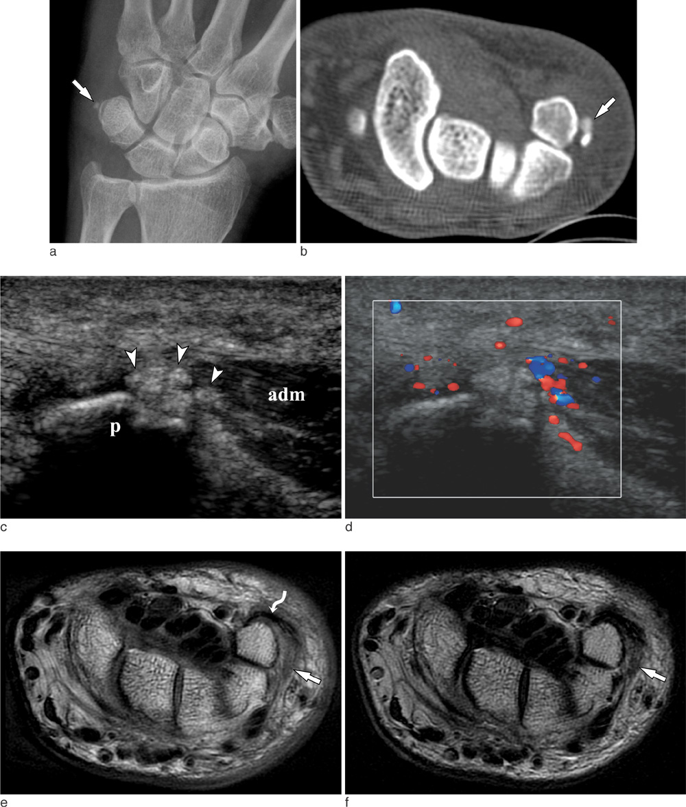
Fig. 3 A 61-year-old man with calcific tendinitis of the abductor digiti minimi. Posteroanterior radiograph (a) and CT (b) of the left wrist show a small calcification (arrow) medial to the pisiform. c. Longitudinal US shows lobulated calcific deposits (arrowheads) in the origin of abductor digiti minimi, adjacent to the pisiform. d. Color Doppler US reveals increased vascularity around the calcific deposits. On axial T1-weighted (e) and T2-weighted (f) images, the calcification is seen as an ill-defined low signal intensity lesion (arrow) in the abductor digiti minimi muscle. Mild soft tissue edema is seen around the calcification. Flexor carpi ulnaris tendon (curved arrow) is indicated. p = pisiform, adm = abductor digiti minimi muscle.
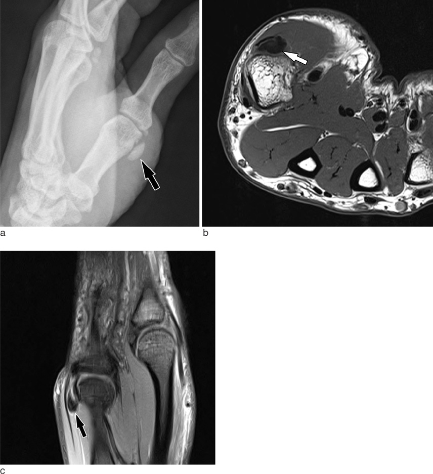
Fig. 4 A 34-year-old man with calcific tendinitis of the abductor pollicis brevis. a. Radiograph of right thumb demonstrates an ovoid calcification (arrow) adjacent to the 1st metacarpal head. Axial T1-weighted (b) and coronal fat-suppressed proton density-weighted (c) MR images show a calcification (arrow) with surrounding edema in the abductor pollicis brevis tendon.
Reference
-
1. Siegal DS, Wu JS, Newman JS, Del Cura JL, Hochman MG. Calcific tendinitis: a pictorial review. Can Assoc Radiol J. 2009. 60:263–272.2. Farin PU, Jaroma H. Sonographic findings of rotator cuff calcifications. J Ultrasound Med. 1995. 14:7–14.3. Lee HS, Lee YH, Sung NK, et al. Sonographic Findings of Calcific Tendinitis around the Hip. J Korean Soc Ultrasound Med. 2005. 24:139–144.4. Shields JS, Chhabra AB, Pannunzio ME. Acute calcific tendinitis of the hand: 2 case reports involving the abductor pollicis brevis. Am J Orthop. 2007. 36:605–607.5. Doumas C, Vazirani RM, Clifford PD, Owens P. Acute calcific periarthritis of the hand and wrist: a series and review of the literature. Emerg Radiol. 2007. 14:199–203.6. Harris AR, McNamara TR, Brault JS, Rizzo M. An unusual presentation of acute calcific tendinitis in the hand. Hand. 2009. 4:81–83.7. Hakozaki M, Iwabuchi M, Konno S, Kikuchi S. Acute calcific tendinitis of the thumb in a child: a case report. Clin Rheumatol. 2007. 26:841–844.8. Woo JH, Lee S, Hong SJ, Song GG. Calcific tendinitis of flexor carpi ulnaris insertion site. J Korean Rheum Assoc. 2010. 17:98–99.9. Klammer G, Iselin LD, Bonel HM, Weber M. Calcific tendinitis of the peroneus longus: case report. Foot Ankle Int. 2011. 32:638–640.
- Full Text Links
-
- Actions
-
Cited
- CITED
-
- Close
- Share
-
- Similar articles
-
- Calcific Tendinitis of Peroneus Longus Tendon (A Case Report)
- Arthroscopic Treatment of Calcific Tendinitis of Subscapularis Tendon: A Case Report
- Acute Calcific Tendinitis of the Abductor Pollicis Brevis in the Hand
- Arthroscopic treatment of chronic calcific tendinitis with intraosseous migration: a case report
- Diagnosis and treatment of calcific tendinitis of the shoulder
