Sacrococcygeal Fetus-in-Fetu Mimicking a Teratoma: A Rare Case with Brain Tissue and an Immature Teratoma Component
- Affiliations
-
- 1Department of Radiology and Research Institute of Radiology, Asan Medical Center, University of Ulsan College of Medicine, Seoul, Korea. chyoon@amc.seoul.kr
- 2Department of Neonatology, Asan Medical Center, University of Ulsan College of Medicine, Seoul, Korea.
- 3Department of Pathology, Asan Medical Center, University of Ulsan College of Medicine, Seoul, Korea.
- KMID: 2097972
- DOI: http://doi.org/10.3348/jksr.2012.67.3.205
Abstract
- Fetus in fetu is a rare, nonviable, malformed parasitic twin, which grows within the body of its partner. It has been known as being almost always anencephalic and rarely reported to have an immature teratoma component. We report a case of a sacrococcygeal fetus-in-fetu with brain tissue seen on both imaging studies and pathologic specimens, containing an immature teratoma component on pathologic examinations. Imaging studies including plain radiography were very helpful for the correct diagnosis.
Figure
-
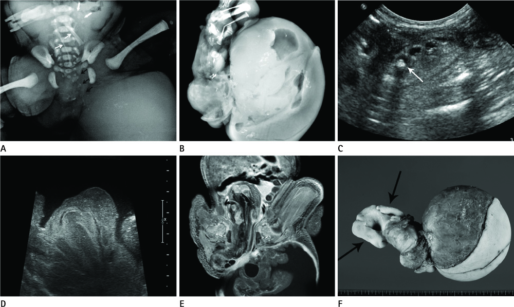
Fig. 1 A sacrococcygeal fetus in fetu in a newborn baby girl. A. Abdominal radiograph shows a large buttock mass containing multiple amorphous calcifications. There are at least two long bones (thin white arrows), vertebral bodies (black arrows), and phalanges-like bones (thick white arrows). B. Specimen radiograph of the excised mass reveals long bones representing the femur, tibia and fibula, short tubular bones representing the feet and hands, and a rounded ilium-like bone. The long bone was accidentally broken during manipulation of the mass. C. The postnatal ultrasonogram shows small contiguous hypoechoic structures with a focal calcification (arrow), which looks like an underdeveloped vertebral column. D. There are convoluted band-like structures representing the brain cortices with cystic areas, using a high resolution linear probe. E. The T1 weighted gadolinium enhanced coronal image shows convoluted band-like structures with cystic areas in the buttock mass. It seems like brain tissue and a venticle. F. Photographs of the gross specimen. There is a large, round head-like structure with two feet (arrows) on the other end. Feet are well developed and covered with skin.
Reference
-
1. Willis RA. The structure of teratomata. J Pathol Bacteriol. 1935. 40:1–36.2. Gonzalez-Crussi F. Extragonadal teratomas. Atlas of tumor pathology. 1982. 2nd ed. Washington, DC: Armed Forces Institute of Pathology;62–79.3. Pourang H, Sarmadi S, Mireskandari SM, Soleimani M, Mollaeian M, Alizadeh H, et al. Twin fetus in fetu with immature teratoma: a case report and review of the literature. Arch Iran Med. 2009. 12:507–510.4. Federici S, Prestipino M, Domenichelli V, Antonellini C, Sciutti R, Dòmini R. Fetus in fetu: report of an additional, well-developed case. Pediatr Surg Int. 2001. 17:483–485.5. Patankar T, Fatterpekar GM, Prasad S, Maniyar A, Mukherji SK. Fetus in fetu: CT appearance--report of two cases. Radiology. 2000. 214:735–737.6. Hoeffel CC, Nguyen KQ, Phan HT, Truong NH, Nguyen TS, Tran TT, et al. Fetus in fetu: a case report and literature review. Pediatrics. 2000. 105:1335–1344.7. Shin JH, Yoon CH, Cho KS, Lim SD, Kim EA, Kim KS, et al. Fetus-in-fetu in the scrotal sac of a newborn infant: imaging, surgical and pathological findings. Eur Radiol. 1999. 9:945–947.8. Hong JH, Kim JH, Kim HJ, Lee IG, Shin JY, Kim DJ. Imaging findings of fetus in fetu: a case report. J Korean Soc Med Ultrasound. 2004. 23:197–201.9. Hong SS, Goo HW, Jung MR, Kim HJ, Kim EA, Kim KS, et al. Fetus in fetu: three-dimensional imaging using multi-detector CT. AJR Am J Roentgenol. 2002. 179:1481–1483.
