Magnetic Resonance Imaging of the Intradural Prepontine Chordoma Mimicking an Epidermoid Cyst: Pathologic Correlation
- Affiliations
-
- 1Department of Radiology, Eulji University Hospital, Daejeon, Korea. midosyu@eulji.ac.kr
- 2Department of Neurosurgery, Eulji University Hospital, Daejeon, Korea.
- KMID: 2097953
- DOI: http://doi.org/10.3348/jksr.2012.67.2.73
Abstract
- Intracranial chordomas, originating from remnants of the primitive notochord, are extradural tumors arising mostly at the sphenooccipital synchondrosis in the clivus. We present an unusual case of intradural chordoma at the prepontine cistern, with parenchymal compressive invasion to the pons. It was excised subtotally, followed by a second operation due to the increasing remnant tumor size during 8 months. A differential diagnosis for intradural chordoma must be considered when the preoperative MRI features are not consistent with an epidermoid cyst if there are multiple fine enhancing lesions on enhanced magnetic resonance images and no bright signal intensity on diffusion-weighted images. This report is concerned with the radiological findings in the intradural chordoma and the differential diagnosis focused on the epidermoid cyst.
MeSH Terms
Figure
-
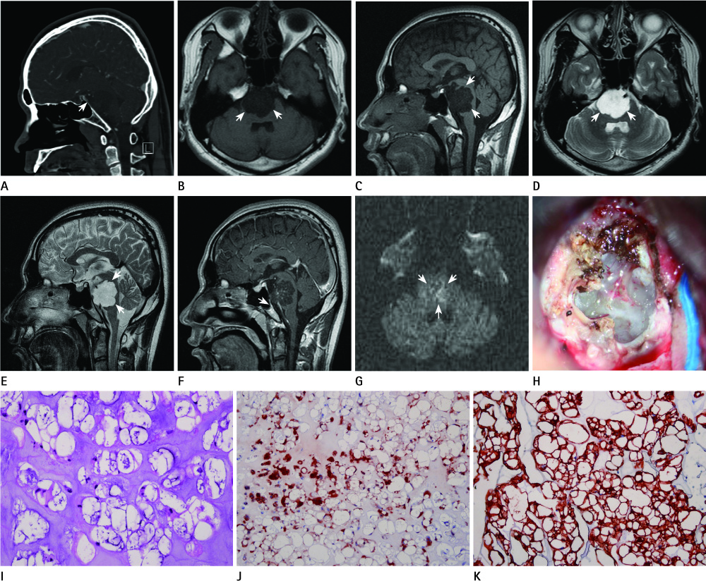
Fig. 1 32-year-old man who has a prepontine cisternal mass. A. Bone setting CT reveals a hypodense prepontine mass with no bony erosion or destruction at the clivus (arrow). B, C. T1-weighted axial (B) and sagittal (C) images show a low signal intensity of the tumor (arrows). D, E. T2-weighted images (D, E) show heterogenous a high signal intensity of the tumor (arrows). F. Most of the mass shows no post-contrast enhancement except some central multiple small reticulonodular heterogenous enhancement. The post-enhancing T1-weighted sagittal image shows the normal clivus (arrow). G. Diffusion-weighted image shows heterogenous subtle increased signal intensity with central decreased signal intensity foci of the tumor (arrows). H. The intraoperative mass is intradurally pale, grayish, gelatinous, glistening, and soft. I. Typical histology of a chordoma with 'physalliferous' eosinophilic cells containing vacuolated cytoplasm, within a pale blue myxoid stroma (H&E stain, × 400). J. The cells are immunopositive for S-100 (× 200). K. The cells are immunopositive for cytokeratin (× 200).
Reference
-
1. Erdem E, Angtuaco EC, Van Hemert R, Park JS, Al-Mefty O. Comprehensive review of intracranial chordoma. Radiographics. 2003; 23:995–1009.2. Mapstone TB, Kaufman B, Ratcheson RA. Intradural chordoma without bone involvement: nuclear magnetic resonance (NMR) appearance. Case report. J Neurosurg. 1983; 59:535–537.3. Rotondo M, Natale M, Mirone G, Cirillo M, Conforti R, Scuotto A. A rare symptomatic presentation of ecchordosis physaliphora: neuroradiological and surgical management. J Neurol Neurosurg Psychiatry. 2007; 78:647–649.4. Meyer JE, Oot RF, Lindfors KK. CT appearance of clival chordomas. J Comput Assist Tomogr. 1986; 10:34–38.5. Oot RF, Melville GE, New PF, Austin-Seymour M, Munzenrider J, Pile-Spellman J, et al. The role of MR and CT in evaluating clival chordomas and chondrosarcomas. AJR Am J Roentgenol. 1988; 151:567–575.6. Nishigaya K, Kaneko M, Ohashi Y, Nukui H. Intradural retroclival chordoma without bone involvement: no tumor regrowth 5 years after operation. Case report. J Neurosurg. 1998; 88:764–768.7. Ito E, Saito K, Nagatani T, Ishiyama J, Terada K, Yoshida M, et al. Intradural cranial chordoma. World Neurosurg. 2010; 73:194–197. discussion e31.8. Gualdi GF, Di Biasi C, Trasimeni G, Pingi A, Vignati A, Maira G. Unusual MR and CT appearance of an epidermoid tumor. AJNR Am J Neuroradiol. 1991; 12:771–772.9. Mehnert F, Beschorner R, Küker W, Hahn U, Nägele T. Retroclival ecchordosis physaliphora: MR imaging and review of the literature. AJNR Am J Neuroradiol. 2004; 25:1851–1855.10. Soo MY, Ng T, Gomes L, Da Cruz M, Dexter M. Skull base chordoid meningioma: imaging features and pathology. Australas Radiol. 2004; 48:233–236.
- Full Text Links
-
- Actions
-
Cited
- CITED
-
- Close
- Share
-
- Similar articles
-
- Imaging Findings of a Nonenhancing Intradural Paramedian Chordoma Mimicking an Epidermoid Cyst
- Clival Cystic Chordoma in Children with Confused Magnetic Resonance Imaging
- Prepontine Chordoma without Bone Involvement: Case Reort
- Epidermoid Cyst after Groin Flap Mimicking Malignancy
- A Rare Case of Thoracic Intradural Epidermoid Cyst after Spinal Cord Stimulator Insertion: A Case Report
