J Korean Soc Surg Hand.
2016 Sep;21(3):167-172. 10.12790/jkssh.2016.21.3.167.
Epidermoid Cyst after Groin Flap Mimicking Malignancy
- Affiliations
-
- 1Department of Orthopaedic Surgery, Wonkwang University School of Medicine, Iksan, Korea. kanghongje@hanmail.net
- 2Department of Orthopaedic Surgery, Bumin Hospital, Busan, Korea.
- KMID: 2353823
- DOI: http://doi.org/10.12790/jkssh.2016.21.3.167
Abstract
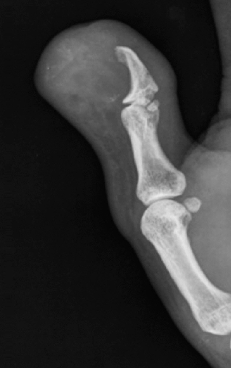
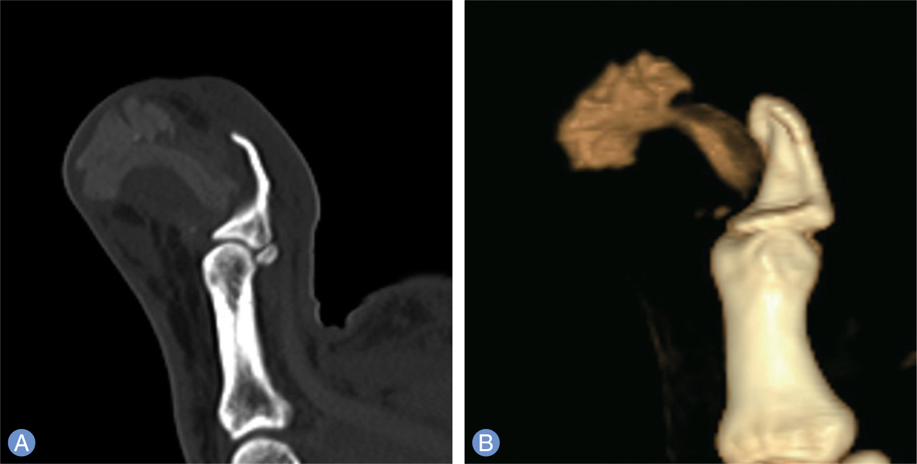
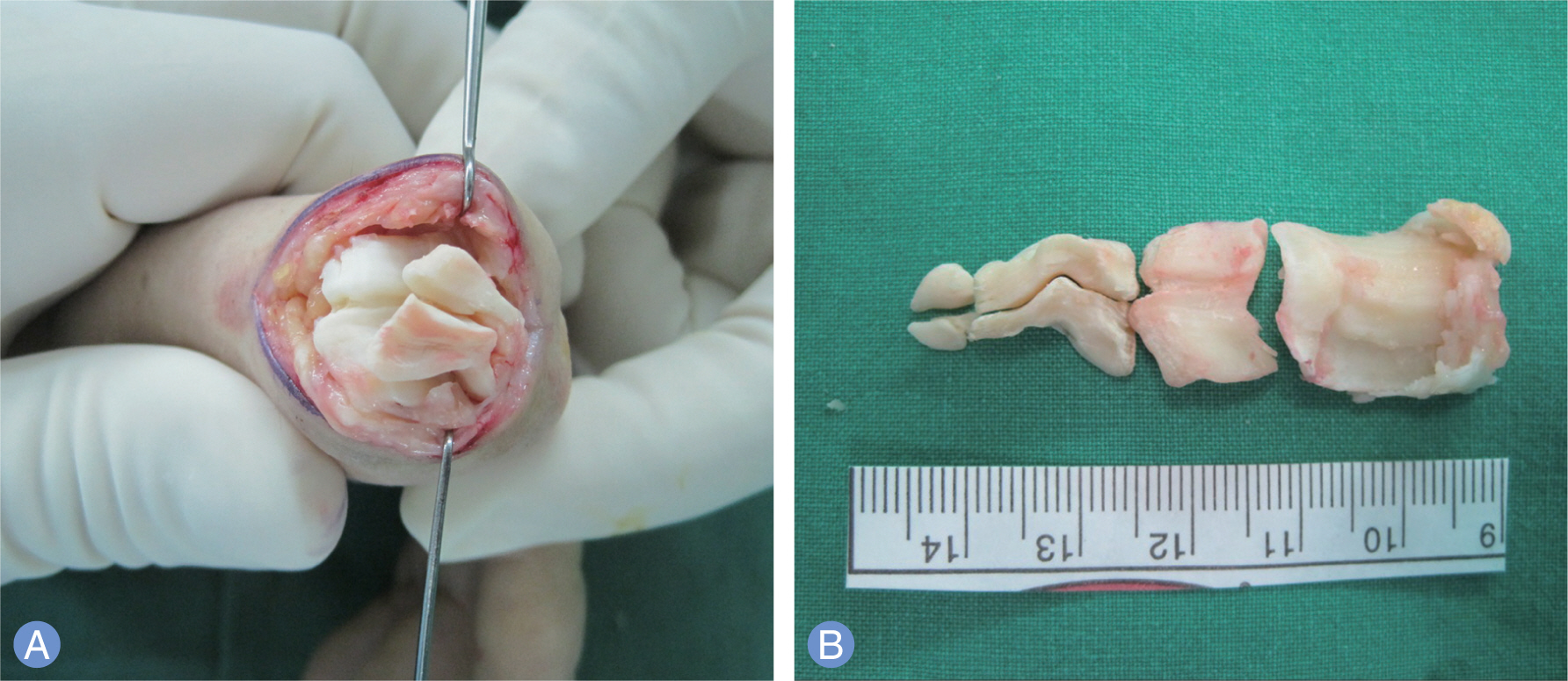
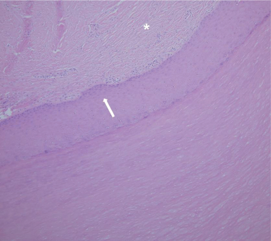
- Epidermoid cyst is a benign tumor containing a layer composed by stratified squamous epithelium and filled with keratin. The epidermoid cyst after soft tissue damage such as bite, laceration could be caused by implantation of epidermal cells. There are reports of epidermoid cyst rarely occurred after surgical procedures such as bone graft or spine puncture. However, the report of epidermoid cyst associated with flap in the hand is very rare. We experienced such epidermoid cyst after the groin flap mimicking malignancy in the distal phalanx of the thumb. We found calcified mass with bony erosions in radiologic findings and heterotrophic signals and partial necrosis in magnetic resonance imaging that suggested malignancy. However, it was pathologically diagnosed as an epidermoid cyst. Therefore, we report the case and literature review.
Keyword
MeSH Terms
Figure
Reference
-
1. Lincoski CJ, Bush DC, Millon SJ. Epidermoid cysts in the hand. J Hand Surg Eur Vol. 2009; 34:792–6.
Article2. Wong CH, Chow L, Yen CH, Ho PC, Yip R, Hung LK. Uncommon hand tumours. Hand Surg. 2001; 6:67–80.
Article3. Penny I, Hooper G. Nail retention cyst: a late complication of digital reconstruction using pedicle flaps. J Hand Surg Br. 1989; 14:345–6.
Article4. Ramakrishna Y, Sudhindra B, Munshi AK. Post-traumatic epidermoid inclusion cyst in the chin region. J Clin Pediatr Dent. 2009; 33:251–2.
Article5. Hwang SM, Kim HI, Ahn SM, Lim KR. Multiple epidermal inclusion cysts in previous bone graft site of the thumb: a case report. J Korean Soc Surg Hand. 2011; 16:247–50.6. Park MR, Lee KH, Lim KM. Inclusion cyst after flap surgery for thumb degloving injury: report of one case. J Korean Soc Surg Hand. 1997; 2:154–7.7. Van Tongel A, De Paepe P, Berghs B. Epidermoid cyst of the phalanx of the finger caused by nail biting. J Plast Surg Hand Surg. 2012; 46:450–1.
Article8. Simon K, Leithner A, Bodo K, Windhager R. Intraosseous epidermoid cysts of the hand skeleton: a series of eight patients. J Hand Surg Eur Vol. 2011; 36:376–8.
Article9. Fisher AR, Mason PH, Wagenhals KS. Ruptured plantar epidermal inclusion cyst. AJR Am J Roentgenol. 1998; 171:1709–10.
Article


