Glioblastoma Multiforme in the Trigone of the Lateral Ventricle: A Case Report
- Affiliations
-
- 1Department of Radiology, Gil Hospital, Gachon University, Korea. h2y@gilhospital.com
- KMID: 2097909
- DOI: http://doi.org/10.3348/jksr.2010.63.5.409
Abstract
- Glioblastoma multiforme (GBM) within the lateral ventricle is relatively rare and it is predominantly found in the frontal horn or body of the ventricle. It is highly unusual to find a GBM in the trigone of the lateral ventricle. We present here a very rare location of a GBM (the trigone of the lateral ventricle).
MeSH Terms
Figure
-
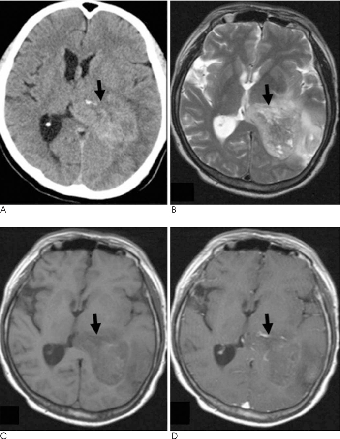
Fig. 1 A 41-year-old man presenting with an episode of headache, nausea and vomiting for three days prior to visit. A. Non-contrast CT shows a poorly defined heterogeneous high attenuated mass within trigone of left lateral ventricle. At axial (B) T2 - & (C) T1-weighted MR images shows heterogeneous signal intensity mass within trigone of left lateral ventricle with local expansion of the ventricle. Mass shows hyperintensity, compared with gray matter on T2 - & T1-weighted MR images. D. Axial gadolinium-enhanced T1-weighted MR images demonstrate a minimally enhancing mass within trigone of left lateral ventricle. Non enhancing hyper and hypointense lesion on T2WI and T1WI within tumor is possibility of intratumoral necrosis (black arrow).
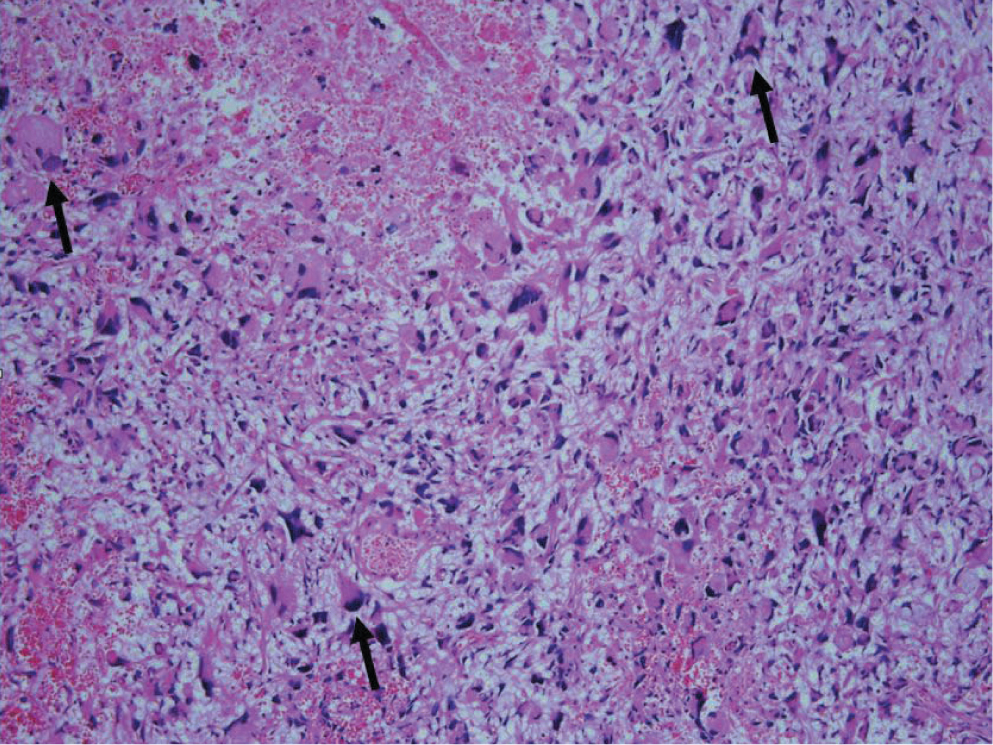
Fig. 2 The mass shows multiple bizarre cells with plump eosinophilic cytoplasm and frequent necrosis (black arrows). (Hematoxylin & eosin stain, × 100)
Reference
-
1. Kleihues P CW. Pathology and genetics of tumors of the nervous system. Lyon: IARC Press;2000.2. Kim YJ, Lee SK, Cho MK. Intraventricular glioblastoma multiforme with previous history of intracerebral hemorrhage : a case report. J Korean Neurosurg Soc. 2008; 44:405–408.3. Park P, Choksi VR, Gala VC, Kaza AR, Murphy HS, Ramnath S. Well-circumscribed, minimally enhancing glioblastoma multiforme of the trigone: a case report and review of the literature. AJNR Am J Neuroradiol. 2005; 26:1475–1478.4. Pendl G, Ozturk E, Haselsberger K. Surgery of tumours of the lateral ventricle. Acta Neurochir (Wien). 1992; 116:128–136.5. Hambly NM, Farrell MA, Scanlon TG, McErlean A, Kavanagh EC. Case report. Glioblastoma multiforme presenting as a haemorrhagic minimally enhancing mass of the trigone. Br J Radiol. 2009; 82:E204–E207.6. Duong H, Sarazin L, Bourgouin P, Vezina JL. Magnetic resonance imaging of lateral ventricular tumours. Can Assoc Radiol J. 1995; 46:434–442.7. Andoh T, Shinoda J, Miwa Y, Hirata T, Sakai N, Yamada H, et al. Tumors at the trigone of the lateral ventricle--clinical analysis of eight cases. Neurol Med Chir (Tokyo). 1990; 30:676–684.8. Tien RD. Intraventricular mass lesions of the brain: CT and MR findings. AJR Am J Roentgenol. 1991; 157:1283–1290.9. Morrison G, Sobel DF, Kelley WM, Norman D. Intraventricular mass lesions. Radiology. 1984; 153:435–442.
- Full Text Links
-
- Actions
-
Cited
- CITED
-
- Close
- Share
-
- Similar articles
-
- Intractable Headache Related to Intraventricular Glioblastoma: A Case Report and Literature Review
- A Case of Meningioma Compatible with Metastatic Glioblastoma Multiforme
- Two Cases of Glioblastoma Multiforme in Children
- A Case of Multicentric Glioblastoma Multiforme
- Congenital Glioblastoma Multiforme: A Case Report
