Undifferentiated carcinoma of the pancreas with osteoclast-like giant cells
- Affiliations
-
- 1Division of Hepatico Biliary Pancreatic Surgery, Department of Surgery, Chonnam National University Medical School, Gwangju, Korea. chcho@jnu.ac.kr
- 2Department of Surgery, Mokpo Christian Hospital, Mokpo, Korea.
- 3Department of Radiology, Chonnam National University Medical School, Gwangju, Korea.
- 4Department of Pathology, Chonnam National University Medical School, Gwangju, Korea.
- KMID: 2096670
- DOI: http://doi.org/10.4174/jkss.2011.81.2.146
Abstract
- Undifferentiated carcinoma with osteoclast-like giant cells is a rare neoplasm of the exocrine pancreas. Some similar cases have been reported, but the histogenesis of these tumors varies and is controversial. We report here on a case of undifferentiated carcinoma of the pancreas with osteoclast-like giant cells. A 77-year old woman presented with abdominal pain and anorexia. Abdominal computed tomography and magnetic resonance imaging showed an approximately 10 x 5 cm highly attenuated mass arising from the tail of the pancreas and invading the spleen and adjacent bowel loop. The initial impression was a malignant endocrine tumor or solid-pseudopapillary tumor of the pancreas. The patient underwent a distal pancreatectomy with splenectomy and left hemicolectomy. The histopathology and immunohistochemistry helped make the diagnosis that of an undifferentiated carcinoma with osteoclast-like giant cells of the pancreas.
MeSH Terms
Figure
-
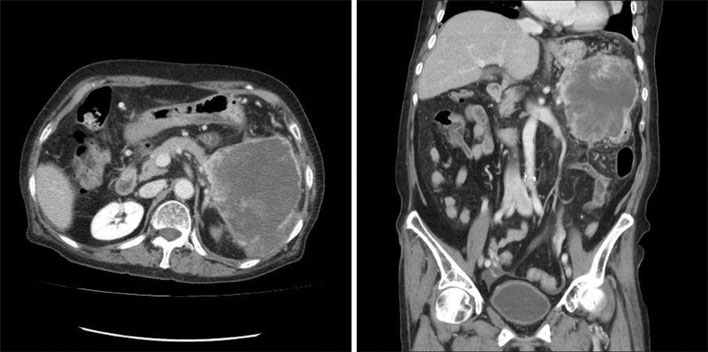
Fig. 1 Abdominal computed tomography (CT) findings. Abdominal CT scan reveals about a 12 × 10 cm-sized heterogenous enhanced mass arising from the tail of the pancreas. The mass has invaded the spleen and the adjacent bowel loop.
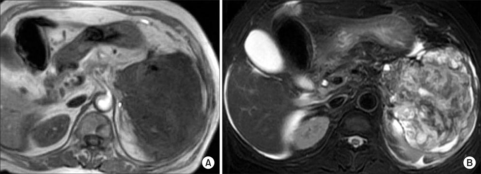
Fig. 2 Abdominal magnetic resonance imaging findings. (A) The T1-weighted image shows a 10 × 5 cm, low signal intensity mass with invasion into the spleen. (B) The T2-weighted image shows heterogenous high signal intensity with multifocal cystic lesions.
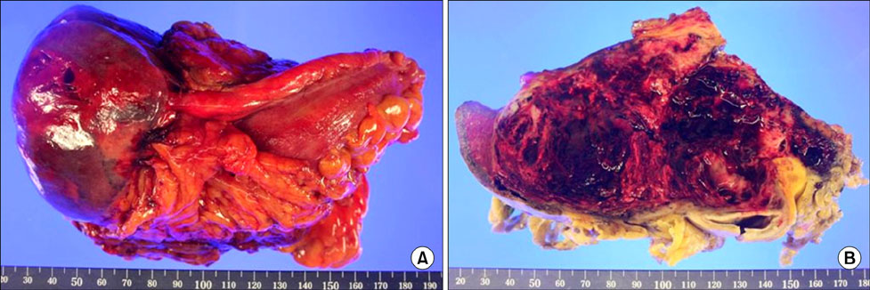
Fig. 3 Gross findings of undifferentiated carcinoma with osteoclast-like giant cells of the pancreas. Gross pathologic examination reveals a 14 × 7.7 cm-sized mass in the pancreatic tail. The cut surface of the tumor is yellowish-white, and the tumor shows signs of hemorrhage and fibrosis.
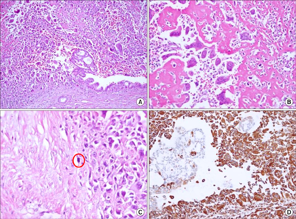
Fig. 4 Microscopic findings. Histologically, the tumor was composed of two major cell types: atypical mononuclear round cells and abundant osteoclast-like multinucleated giant cells with central nucleoli (A, H&E, ×100; B, H&E, ×200). Many mitotic figures are present (C, red circle) (H&E, ×200). The immunohistochemical findings show positive staining by vimentin (D, ×200).
Cited by 1 articles
-
Undifferentiated carcinoma with osteoclast-like giant cells of the pancreas: An individual participant data meta-analysis
Adam Mylonakis, Tatiana S. Driva, Panagis Lykoudis, Maximos Frountzas, Nikolaos Machairas, Dimitrios Tsapralis, Konstantinos G. Toutouzas, Dimitrios Schizas
Ann Hepatobiliary Pancreat Surg. 2024;28(2):125-133. doi: 10.14701/ahbps.23-161.
Reference
-
1. Molberg KH, Heffess C, Delgado R, Albores-Saavedra J. Undifferentiated carcinoma with osteoclast-like giant cells of the pancreas and periampullary region. Cancer. 1998. 82:1279–1287.2. Bauditz J, Rudolph B, Wermke W. Osteoclast-like giant cell tumors of the pancreas and liver. World J Gastroenterol. 2006. 12:7878–7883.3. Moon YH, Chu YC, Kim KR. Giant cell carcinoma of the pancreas of the osteoclastic type associated with a mucinous cystadenocarcinoma. J Korean Surg Soc. 1989. 36:546–551.4. Verbeke CS, Menon KV. Osteoclast-like giant cell tumour of the pancreas: an undifferentiated carcinoma of duct epithelial origin. Pancreatology. 2006. 6:254.5. Sakai Y, Kupelioglu AA, Yanagisawa A, Yamaguchi K, Hidaka E, Matsuya S, et al. Origin of giant cells in osteoclast-like giant cell tumors of the pancreas. Hum Pathol. 2000. 31:1223–1229.6. Nai GA, Amico E, Gimenez VR, Guilmar M. Osteoclast-like giant cell tumor of the pancreas associated with mucus-secreting adenocarcinoma. Case report and discussion of the histogenesis. Pancreatology. 2005. 5:279–284.7. Shiozawa M, Imada T, Ishiwa N, Rino Y, Hasuo K, Takanashi Y, et al. Osteoclast-like giant cell tumor of the pancreas. Int J Clin Oncol. 2002. 7:376–380.8. Oehler U, Jürs M, Klöppel G, Helpap B. Osteoclast-like giant cell tumour of the pancreas presenting as a pseudocyst-like lesion. Virchows Arch. 1997. 431:215–218.9. Gao L, Li ZS, Jin ZD, Man XH, Zhang MH, Zhu MH. Undifferentiated carcinoma with osteoclast-like giant cells of the pancreas diagnosed by endoscopic ultrasonography-guided fine-needle aspiration. Chin Med J (Engl). 2009. 122:1598–1600.10. Goldberg RD, Michelassi F, Montag AG. Osteoclast-like giant cell tumor of the pancreas: immunophenotypic similarity to giant cell tumor of bone. Hum Pathol. 1991. 22:618–622.
- Full Text Links
-
- Actions
-
Cited
- CITED
-
- Close
- Share
-
- Similar articles
-
- A Case of Undifferentiated Carcinoma with Osteoclast-Like Giant Cells in the Pancreas
- Undifferentiated Carcinoma with Osteoclast-Like Giant Cells of the Pancreas Mimicking a Predominantly Calcified Mass: Case Report and Literature Review
- Undifferentiated Gallbladder Carcinoma with Osteoclast-like Giant Cells: A Case Report
- A case of undifferentiated carcinoma with osteoclast-like giant cells of the pancreas
- Undifferentiated Pancreatic Carcinoma with Osteoclast-like Giant Cells: A Case Report
