F18-fluorodeoxyglucose-positron emission tomography and computed tomography is not accurate in preoperative staging of gastric cancer
- Affiliations
-
- 1Department of Surgery, Hanyang University College of Medicine, Seoul, Korea. sjkwon@hanyang.ac.kr
- 2Department of Nuclear Medicine, Hanyang University College of Medicine, Seoul, Korea.
- 3Department of Diagnostic Radiology, Hanyang University College of Medicine, Seoul, Korea.
- KMID: 2096663
- DOI: http://doi.org/10.4174/jkss.2011.81.2.104
Abstract
- PURPOSE
To investigate the clinical benefits of F18-fluorodeoxyglucose-positron emission tomography and computed tomography (18F-FDG-PET/CT) over multi-detector row CT (MDCT) in preoperative staging of gastric cancer.
METHODS
FDG-PET/CT and MDCT were performed on 78 patients with gastric cancer pathologically diagnosed by endoscopy. The accuracy of radiologic staging retrospectively was compared to pathologic result after curative resection.
RESULTS
Primary tumors were detected in 51 (65.4%) patients with 18F-FDG-PET/CT, and 47 (60.3%) patients with MDCT. Regarding detection of lymph node metastasis, the sensitivity of FDG-PET/CT was 51.5% with an accuracy of 71.8%, whereas those of MDCT were 69.7% and 69.2%, respectively. The sensitivity of 18F-FDG-PET/CT for a primary tumor with signet ring cell carcinoma was lower than that of 18F-FDG-PET/CT for a primary tumor with non-signet ring cell carcinoma (35.3% vs. 73.8%, P < 0.01).
CONCLUSION
Due to its low sensitivity, 18F-FDG-PET/CT alone shows no definite clinical benefit for prediction of lymph node metastasis in preoperative staging of gastric cancer.
Keyword
MeSH Terms
Figure
-
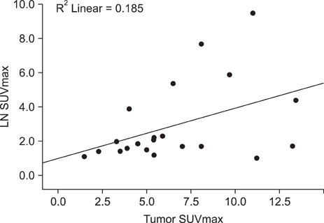
Fig. 1 Correlation analysis curve between primary tumor maximum standardized uptake value (SUVmax) and lymph node (LN) SUVmax.
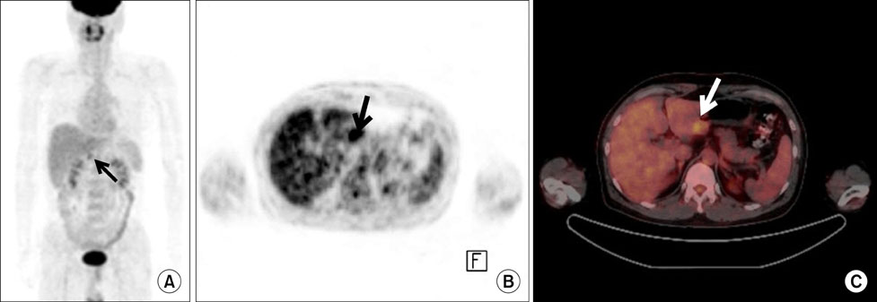
Fig. 2 Early gastric cancer in a 47-year-old man. Miaximum intensity projection image of (A) positron emission tomography (PET) and (B) axial PET scan demonstrate focal hypermetabolic lesion in gastric antrum (black arrow) with maximum standardized uptake value (SUVmax) of 4.3. (C) PET/CT scan shows focal hypermtabolic intraluminal mass in posterior wall of gastric antrum (white arrow).
Cited by 1 articles
-
Does Positron Emission Tomography-Computed Tomography Maximum Standardized Uptake Value Predict Survival in Surgically Resected Gastric Cancer?
Hyun Joo Song
Korean J Gastroenterol. 2014;63(6):333-334. doi: 10.4166/kjg.2014.63.6.333.
Reference
-
1. Parkin DM, Pisani P, Ferlay J. Global cancer statistics. CA Cancer J Clin. 1999. 49:33–64.2. Bae JM, Jung KW, Won YJ. Estimation of cancer deaths in Korea for the upcoming years. J Korean Med Sci. 2002. 17:611–615.3. Fujii K, Isozaki H, Okajima K, Nomura E, Niki M, Sako S, et al. Clinical evaluation of lymph node metastasis in gastric cancer defined by the fifth edition of the TNM classification in comparison with the Japanese system. Br J Surg. 1999. 86:685–689.4. Cervantes A, Roselló S, Roda D, Rodríguez-Braun E. The treatment of advanced gastric cancer: current strategies and future perspectives. Ann Oncol. 2008. 19:Suppl 5. v103–v107.5. Nomura S, Kaminishi M. Surgical treatment of early gastric cancer. Dig Surg. 2007. 24:96–100.6. Woo SK, Kim S, Kim TU, Lee JW, Kim GH, Choi KU, et al. Investigation of the association between CT detection of early gastric cancer and ultimate histology. Clin Radiol. 2008. 63:1236–1244.7. Kumano S, Murakami T, Kim T, Hori M, Iannaccone R, Nakata S, et al. T staging of gastric cancer: role of multi-detector row CT. Radiology. 2005. 237:961–966.8. Kim HJ, Kim AY, Oh ST, Kim JS, Kim KW, Kim PN, et al. Gastric cancer staging at multi-detector row CT gastrography: comparison of transverse and volumetric CT scanning. Radiology. 2005. 236:879–885.9. Bar-Shalom R, Yefremov N, Guralnik L, Gaitini D, Frenkel A, Kuten A, et al. Clinical performance of PET/CT in evaluation of cancer: additional value for diagnostic imaging and patient management. J Nucl Med. 2003. 44:1200–1209.10. Cohade C, Osman M, Leal J, Wahl RL. Direct comparison of (18)F-FDG PET and PET/CT in patients with colorectal carcinoma. J Nucl Med. 2003. 44:1797–1803.11. Lardinois D, Weder W, Hany TF, Kamel EM, Korom S, Seifert B, et al. Staging of non-small-cell lung cancer with integrated positron-emission tomography and computed tomography. N Engl J Med. 2003. 348:2500–2507.12. Lee MC, Oh SW, Chung JK, Lee DS. The current status and future perspectives of nuclear medicine in Korea. Nucl Med Mol Imaging. 2010. 44:95–101.13. Kim AY, Han JK, Seong CK, Kim TK, Choi BI. MRI in staging advanced gastric cancer: is it useful compared with spiral CT? J Comput Assist Tomogr. 2000. 24:389–394.14. Dorfman RE, Alpern MB, Gross BH, Sandler MA. Upper abdominal lymph nodes: criteria for normal size determined with CT. Radiology. 1991. 180:319–322.15. Sohn KM, Lee JM, Lee SY, Ahn BY, Park SM, Kim KM. Comparing MR imaging and CT in the staging of gastric carcinoma. AJR Am J Roentgenol. 2000. 174:1551–1557.16. Sugano H, Nakamura K, Kato Y. Pathological studies of human gastric cancer. Acta Pathol Jpn. 1982. 32:Suppl 2. 329–347.17. Hopper KD, Singapuri K, Finkel A. Body CT and oncologic imaging. Radiology. 2000. 215:27–40.18. Israel O, Mor M, Gaitini D, Keidar Z, Guralnik L, Engel A, et al. Combined functional and structural evaluation of cancer patients with a hybrid camera-based PET/CT system using (18)F-FDG. J Nucl Med. 2002. 43:1129–1136.19. Weber DA, Ivanovic M. Correlative image registration. Semin Nucl Med. 1994. 24:311–323.20. Sim SH, Kim YJ, Oh DY, Lee SH, Kim DW, Kang WJ, et al. The role of PET/CT in detection of gastric cancer recurrence. BMC Cancer. 2009. 9:73.21. Kim SK, Kang KW, Lee JS, Kim HK, Chang HJ, Choi JY, et al. Assessment of lymph node metastases using 18F-FDG PET in patients with advanced gastric cancer. Eur J Nucl Med Mol Imaging. 2006. 33:148–155.22. Chen J, Cheong JH, Yun MJ, Kim J, Lim JS, Hyung WJ, et al. Improvement in preoperative staging of gastric adenocarcinoma with positron emission tomography. Cancer. 2005. 103:2383–2390.23. Minami M, Kawauchi N, Itai Y, Niki T, Sasaki Y. Gastric tumors: radiologic-pathologic correlation and accuracy of T staging with dynamic CT. Radiology. 1992. 185:173–178.24. Fukuya T, Honda H, Hayashi T, Kaneko K, Tateshi Y, Ro T, et al. Lymph-node metastases: efficacy for detection with helical CT in patients with gastric cancer. Radiology. 1995. 197:705–711.25. Shimizu K, Ito K, Matsunaga N, Shimizu A, Kawakami Y. Diagnosis of gastric cancer with MDCT using the water-filling method and multiplanar reconstruction: CT-histologic correlation. AJR Am J Roentgenol. 2005. 185:1152–1158.26. Gore RM. Upper gastrointestinal tract tumours: diagnosis and staging strategies. Cancer Imaging. 2005. 5:95–98.27. Rohren EM, Turkington TG, Coleman RE. Clinical applications of PET in oncology. Radiology. 2004. 231:305–332.28. Mönig SP, Zirbes TK, Schröder W, Baldus SE, Lindemann DG, Dienes HP, et al. Staging of gastric cancer: correlation of lymph node size and metastatic infiltration. AJR Am J Roentgenol. 1999. 173:365–367.29. Kwee RM, Kwee TC. Imaging in assessing lymph node status in gastric cancer. Gastric Cancer. 2009. 12:6–22.
- Full Text Links
-
- Actions
-
Cited
- CITED
-
- Close
- Share
-
- Similar articles
-
- 18F-2-Deoxy-2-Fluoro-D-Glucose Positron Emission Tomography: Computed Tomography for Preoperative Staging in Gastric Cancer Patients
- Transient ¹â¸F-Fluorodeoxyglucose Activity on PET/CT of Herniation Pit in Thyroid Cancer Patient: A Case Report
- Measuring tumor metabolic heterogeneity on positron emission tomography: utility in cervical cancer
- Fluorine-18 Fluorodeoxyglucose Positron Emission Tomography/Computed Tomography Findings of Post Traumatic Lymphangioma in a Young Adult Male
- Fluorodeoxyglucose-positron emission tomography/computed tomography imaging of squamous cell carcinoma arising in a meningomyelocele
