Transient ¹â¸F-Fluorodeoxyglucose Activity on PET/CT of Herniation Pit in Thyroid Cancer Patient: A Case Report
- Affiliations
-
- 1Department of Nuclear Medicine, Eulji University Hospital, Daejeon, Korea.
- 2Department of Radiology, The Catholic University of Korea, Daejeon St. Mary's Hospital, Daejeon, Korea. greempark@gmail.com
- KMID: 2392123
- DOI: http://doi.org/10.3348/jksr.2017.77.4.249
Abstract
- A herniation pit is a benign bone pit characterized by its femoral location and surrounding sclerotic margin. Few reports have been issued on the fluorodeoxyglucose (FDG) positron emission tomography findings of herniation pit. Here, we report the unique case of a thyroid cancer patient, having a herniation pit showing transient FDG uptake, which mimicked bone metastasis.
MeSH Terms
Figure
-
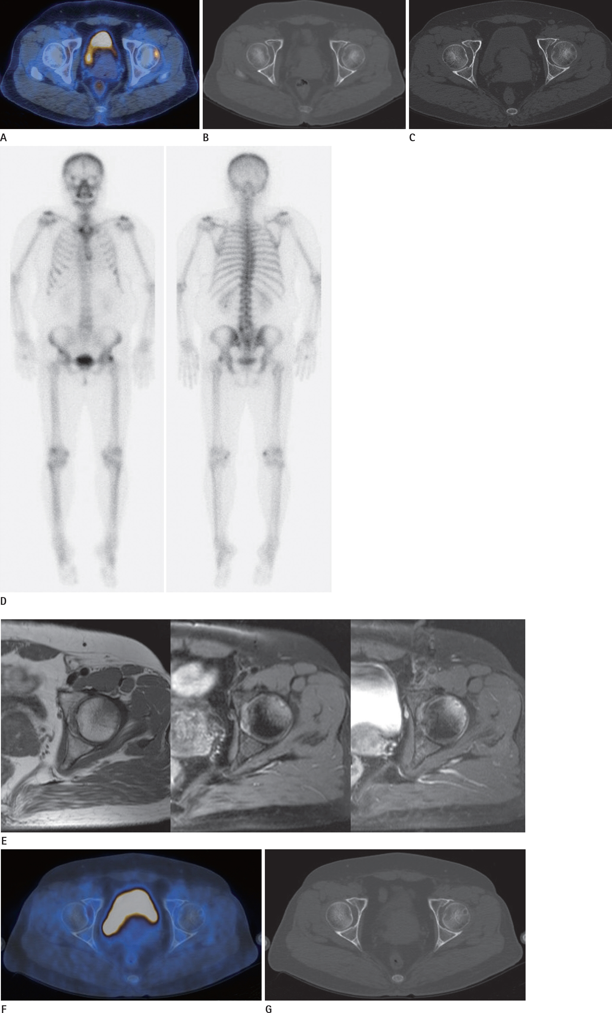
Fig. 1. Transient 18 F-FDG activity of herniation pit, in a 53-year-old woman with thyroid cancer. A. PET/CT fusion image shows a focal hypermetabolism in the left anterolateral femoral head. B. CT bone setting finding, which corresponds to the location of the hypermetabolic lesion, shows no definite abnormality. C. After 3 weeks, axial CT of pelvic bone shows a small hypodense lesion without a sclerotic rim, in the left anterolateral femoral head. D. Bone scan shows focal increased uptake in the left lateral femoral head. CT = computed tomography, FDG = fluorodeoxyglucose, PET = positron emission tomography. Transient 18 F-FDG activity of herniation pit, in a 53-year-old woman with thyroid cancer. E. MR T1-weighted (left), T2-weighted (middle), contrast enhanced T1-weight (right) images. T1 weighted image shows ill defined low SI lesion in the anterosuperior femoral head/neck junction. T2-weighted image shows high SI with adjacent marrow edema. Contrast enhanced T1-weighted image shows focal rim enhancing lesion with mild diffuse enhancement of marrow. F, G. Follow up PET/CT images after 9 months show no FDG uptake (F), and a hypodense lesion with a sclerotic border in the left anterolateral femo-ral head, characteristic of a herniation pit (G). CT = computed tomography, FDG = fluorodeoxyglucose, PET = positron emission tomography, SI = signal intensity
Reference
-
1.Kim JA., Park JS., Jin W., Ryu K. Herniation pits in the femo-ral neck: a radiographic indicator of femoroacetabular im-pingement? Skeletal Radiol. 2011. 40:167–172.
Article2.Sopov V., Fuchs D., Bar-Meir E., Gorenberg M., Groshar D. Clinical spectrum of asymptomatic femoral neck abnormal uptake on bone scintigraphy. J Nucl Med. 2002. 43:484–486.3.Gao ZH., Yin JQ., Ma L., Wang J., Meng QF. Clinical imaging characteristics of herniation pits of the femoral neck. Or-thop Surg. 2009. 1:189–195.
Article4.Yoo SW., Song HC., Oh JR., Kim J., Kang SR., Chong A, et al. Herniation pit mimicking osseous metastasis on 18F-FDG PET/CT in patient with lung cancer. Clin Nucl Med. 2012. 37:682–683.
Article5.Pitt MJ., Graham AR., Shipman JH., Birkby W. Herniation pit of the femoral neck. AJR Am J Roentgenol. 1982. 138:1115–1121.
Article6.Panzer S., Esch U., Abdulazim AN., Augat P. Herniation pits and cystic-appearing lesions at the anterior femoral neck: an anatomical study by MSCT and microCT. Skeletal Radiol. 2010. 39:645–654.7.Kim SH., Yoo HJ., Kang Y., Choi JY., Hong SH. MRI findings of new uptake in the femoral head detected on follow-up bone scans. AJR Am J Roentgenol. 2015. 204:608–614.
Article8.Borody C. Symptomatic herniation pit of the femoral neck: a case report. J Manipulative Physiol Ther. 2005. 28:449–451.
Article9.Min JW., Um SW., Yim JJ., Yoo CG., Han SK., Shim YS, et al. The role of whole-body FDG PET/CT, Tc 99m MDP bone scintig-raphy, and serum alkaline phosphatase in detecting bone metastasis in patients with newly diagnosed lung cancer. J Korean Med Sci. 2009. 24:275–280.
Article10.Gibiezaite S., Ozdemir S., Shuja S., McCook B., Plazarte M., Sheikh-Ali M. Unexpected bone metastases from thyroid cancer. Case Rep Endocrinol. 2015. 2015:434732.
Article
- Full Text Links
-
- Actions
-
Cited
- CITED
-
- Close
- Share
-
- Similar articles
-
- Dual Pathologies of Parathyroid Adenoma and Papillary Thyroid Cancer on Fluorocholine and Fluorodeoxyglucose PET/CT
- â¶â¸Gallium-Arginine-Glycine-Aspartic Acid and ¹â¸F-Fluorodeoxyglucose Positron Emission Tomography/Computed Tomography in Chondroblastic Osteosarcoma of the Skull
- Comparison of Neck CT and ¹â¸F-FDG PET-CT for Making the Preoperative Diagnosis of Lymph Node Metastasis in Papillary Thyroid Cancer
- Characteristics and Detection Rate of Thyroidal Incidentaloma using ¹â¸F-FDG PET-CT
- Role of ¹â¸F-FDG PET-CT in Monitoring the Cyclophosphamide Induced Pulmonary Toxicity in Patients with Breast Cancer: 2 Case Reports
