J Korean Thyroid Assoc.
2012 Nov;5(2):161-162. 10.11106/jkta.2012.5.2.161.
A Case of Killian-Jamieson Diverticulum Mimicking a Thyroid Nodule
- Affiliations
-
- 1Department of Internal Medicine, Soonchunhyang University School of Medicine, Cheonan, Korea. yeojoo@schmc.ac.kr
- KMID: 2095107
- DOI: http://doi.org/10.11106/jkta.2012.5.2.161
Abstract
- No abstract available.
MeSH Terms
Figure
-
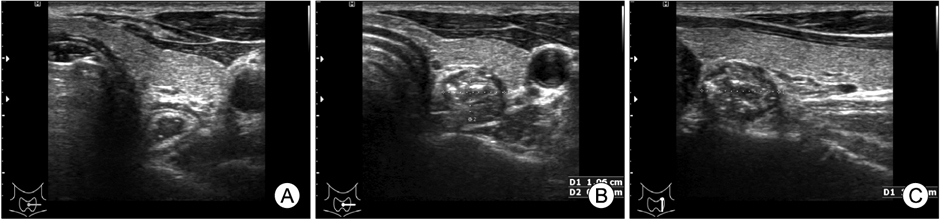
Fig. 1 (A) The axial US scan shows normal esophagus. (B, C) The axial (B) and longitudinal (C) US scan of the left thyroid lobe shows 10.6×8.6×13.4 mm sized low echogenic lesion with some echogenic foci.

Fig. 2 (A, B) The esophagogastroscopy shows a outpouching lesion below the upper esophageal sphincter, containing food material.
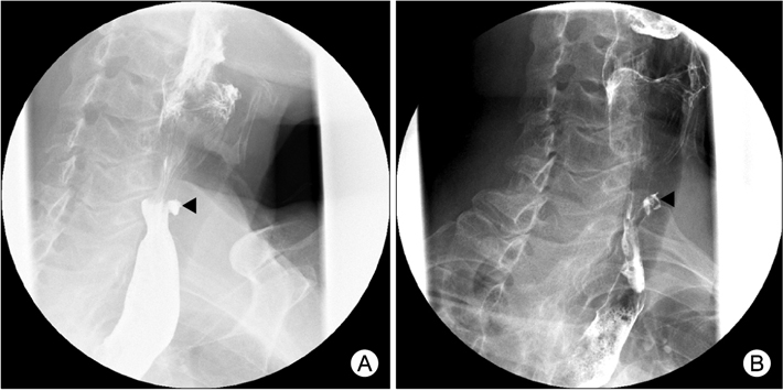
Fig. 3 (A, B) The esophagography confirms a barium-filled sac protruding from the left anterolateral wall of the cervical esophagus (arrowheads).
Reference
-
1. Kim HK, Lee JI, Jang HW, Bae SY, Lee JH, Kim YS, et al. Characteristics of Killian-Jamieson diverticula mimicking a thyroid nodule. Head Neck. 2012. 34(4):599–603.
Article2. Kim SJ, Kim CH. The genetic studies of obsessive-compulsive disorder and its future directions. Yonsei Med J. 2006. 47(4):443–454.
Article3. Yoon HD, Shon HS. Killian-Jamieson diverticulum mimicking a thyroid nodule. Korean J Med. 2005. 68(4):467–468.4. DeFriend DE, Dubbins PA. Sonographic demonstration of a pharyngoesophageal diverticulum. J Clin Ultrasound. 2000. 28(9):485–487.
Article5. Komatsu M, Komatsu T, Inove K. Ultrasonography of Zenker's diverticulum: special reference to differential diagnosis from thyroid nodules. Eur J Ultrasound. 2000. 11(2):123–125.
Article
- Full Text Links
-
- Actions
-
Cited
- CITED
-
- Close
- Share
-
- Similar articles
-
- Killian-Jamieson Diverticula Mimicking Thyroid Nodule on Ultrasound: Radiographic Findings in Two Patients
- Pharyngoesophageal (Killian-Jamieson) Diverticulum Mimicking Thyroid Nodule on Ultrasonography: A Case Report
- Killian-Jamieson diverticulum mimicking a thyroid nodule
- Killian-Jamieson Diverticulum Mimicking a Thyroid Nodule on Ultrasonography: A Case Report
- Killian-Jamieson Diverticula Mimicking a Right Thyroid Nodule on Ultrasonography: A Case Report
