Primary Melanoma of the Breast: A Case Report with Imaging Findings
- Affiliations
-
- 1Department of Radiology and Research Institute of Radiology, Asan Medical Center, University of Ulsan College of Medicine, Seoul, Korea. jhcha@amc.seoul.kr
- 2Department of Pathology, Asan Medical Center, University of Ulsan College of Medicine, Seoul, Korea.
- KMID: 2079562
- DOI: http://doi.org/10.3348/jksr.2015.73.5.287
Abstract
- Primary breast melanoma is extremely rare, and as such, there are no established radiologic findings in the literature. This report describes a case of primary malignant melanoma with mammography, ultrasonography, and magnetic resonance imaging findings. Our case study demonstrates a well-circumscribed heterogeneous rim-enhancing mass, with an internal cystic or necrotic portion seen using three modalities. Thus, although rare, this condition should be included in the differential diagnosis of a well-demarcated heterogeneous breast mass, and further pathological confirmation is needed.
MeSH Terms
Figure
-
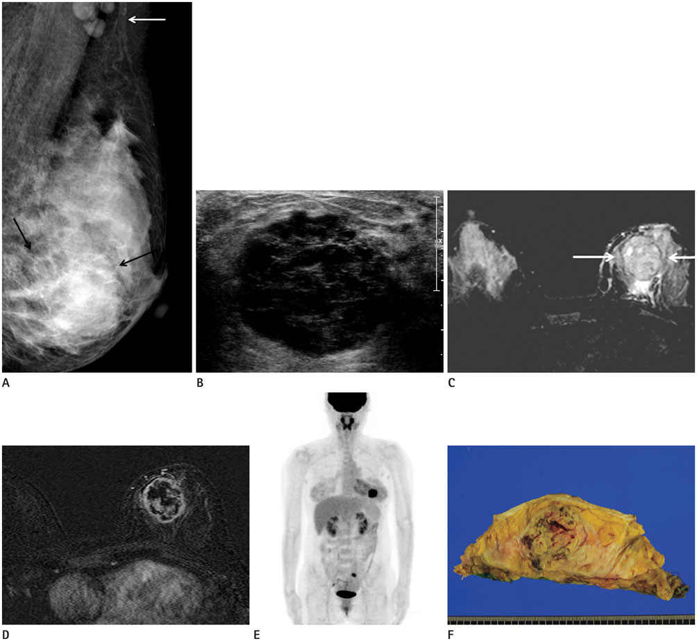
Fig. 1 A 45-year-old woman with a palpable mass in the left breast that she noticed 2 months previously. A. Left medio-lateral oblique showing an approximate 4-cm sized, round, ill-defined, high-density mass (black arrows) in the lower portion of the left breast, and enlarged lymph nodes (white arrow) in the axilla. B. Ultrasonography showing a round, microlobulated, low echoic mass with internal cystic portions in the lower inner quadrant of the left breast. C, D. Magnetic resonance imaging showing a well-defined mass with heterogeneous high signal intensity and a dark rim on T2-weighted image (arrows) (C), and irregular enhancement on the rim and septum after contrast injection (D). E. PET-CT scan shows hypermetabolic uptake in the left breast mass, and no other abnormal hypermetabolic lesion. F. On gross specimen examination, a firm, lobulated, and multifocal necrotic mass (5 × 4 × 4 cm) is revealed. The cut surface is yellowish white and granular, and the overlying skin is not involved. PET-CT = positron emission tomography-CT
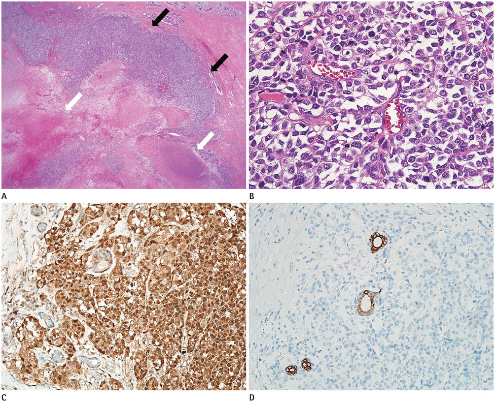
Fig. 2 Microscopic images of the breast mass. A. In the low power view (× 40), hypercellular mass is infiltrating to the adjacent breast parenchyma (black arrows). Multifocal necrosis and hemorrhage are present in the tumor (white arrows). B. In the high power view (× 400), the tumor shows a diffuse, solid growth pattern without ductal differentiation. Tumor cells are highly pleomorphic and have clear cytoplasm and atypical nuclei. Mitoses are frequently seen. Intracellular pigments or granules are not observed. C, D. On immunohistochemical staining (× 400), tumor cells are positive for S-100 protein (C) and HMB45 (not shown), but negative for cytokeratin (D), while the entrapped non-neoplastic ducts are negative for S-100 protein (C) and positive for cytokeratin (D). The overall features support the diagnosis of malignant melanoma.
Reference
-
1. Ravdel L, Robinson WA, Lewis K, Gonzalez R. Metastatic melanoma in the breast: a report of 27 cases. J Surg Oncol. 2006; 94:101–104.2. Bernardo MM, Mascarenhas MJ, Lopes DP. Primary malignant melanoma of the breast. Acta Med Port. 1980; 2:39–43.3. Toombs BD, Kalisher L. Metastatic disease to the breast: clinical, pathologic, and radiographic features. AJR Am J Roentgenol. 1977; 129:673–676.4. Teodorescu EC. Sonography and mammography of primary malignant breast melanoma. Med Ultrason. 2008; 10:55–58.5. Loffeld A, Marsden JR. Management of melanoma metastasis to the breast: case series and review of the literature. Br J Dermatol. 2005; 152:1206–1210.6. Bassi F, Gatti G, Mauri E, Ballardini B, De Pas T, Luini A. Breast metastases from cutaneous malignant melanoma. Breast. 2004; 13:533–535.7. He Y, Mou J, Luo D, Gao B, Wen Y. Primary malignant melanoma of the breast: a case report and review of the literature. Oncol Lett. 2014; 8:238–240.8. Ohsie SJ, Sarantopoulos GP, Cochran AJ, Binder SW. Immunohistochemical characteristics of melanoma. J Cutan Pathol. 2008; 35:433–444.
- Full Text Links
-
- Actions
-
Cited
- CITED
-
- Close
- Share
-
- Similar articles
-
- A Case of Malignant Melanoma Presenting as a Breast Mass
- Recurrent Primary Pleomorphic Liposarcoma of the Breast: A Case Report with Imaging Findings
- Primary cutaneous malignant melanoma of the breast
- Bladder Cancer Metastasis to the Breast in a Male Patient: Imaging Findings on Mammography and Ultrasonography
- Fine Needle Aspiration Cytology of Metastatic Melanoma in the Breast: A Case Report
