A Study to Compare the Radiation Absorbed Dose of the C-arm Fluoroscopic Modes
- Affiliations
-
- 1Department of Anesthesiology and Pain Medicine, Konkuk University Medical Center, Seoul, Korea. painfree@kuh.ac.kr
- KMID: 2074014
- DOI: http://doi.org/10.3344/kjp.2011.24.4.199
Abstract
- BACKGROUND
Although many clinicians know about the reducing effects of the pulsed and low-dose modes for fluoroscopic radiation when performing interventional procedures, few studies have quantified the reduction of radiation-absorbed doses (RADs). The aim of this study is to compare how much the RADs from a fluoroscopy are reduced according to the C-arm fluoroscopic modes used.
METHODS
We measured the RADs in the C-arm fluoroscopic modes including 'conventional mode', 'pulsed mode', 'low-dose mode', and 'pulsed + low-dose mode'. Clinical imaging conditions were simulated using a lead apron instead of a patient. According to each mode, one experimenter radiographed the lead apron, which was on the table, consecutively 5 times on the AP views. We regarded this as one set and a total of 10 sets were done according to each mode. Cumulative exposure time, RADs, peak X-ray energy, and current, which were viewed on the monitor, were recorded.
RESULTS
Pulsed, low-dose, and pulsed + low-dose modes showed significantly decreased RADs by 32%, 57%, and 83% compared to the conventional mode. The mean cumulative exposure time was significantly lower in the pulsed and pulsed + low-dose modes than in the conventional mode. All modes had pretty much the same peak X-ray energy. The mean current was significantly lower in the low-dose and pulsed + low-dose modes than in the conventional mode.
CONCLUSIONS
The use of the pulsed and low-dose modes together significantly reduced the RADs compared to the conventional mode. Therefore, the proper use of the fluoroscopy and its C-arm modes will reduce the radiation exposure of patients and clinicians.
MeSH Terms
Figure
-
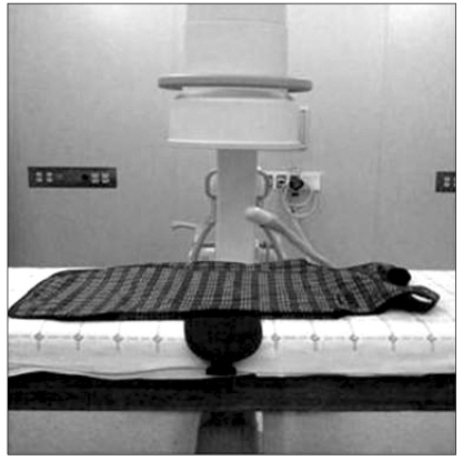
Fig. 1 The lead apron (0.85 mm lead equivalent thickness) was on the table and the X-ray tube was positioned below the apron and the detector above the apron.
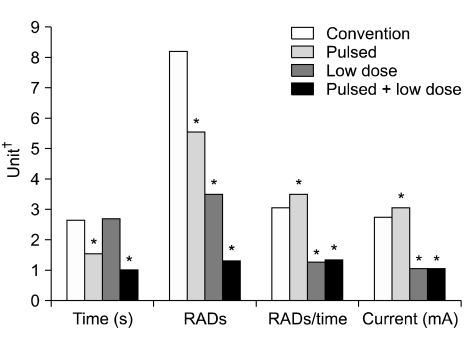
Fig. 2 Comparison of time, radiation-absorbed doses (RADs), mean RADs/mean time and current (mA) among modes by graphs. Pulsed fluoroscopic mode of 15 frames per second was used. *P < 0.05. †Unit is expressed as second in Time, mRADs/cm2 in RADs, mRADs/cm2 · second in RADs/Time, and mA in Current.

Fig. 3 Comparison of peak X-ray energy (kVp) among modes by graphs. Pulsed fluoroscopic mode of 15 frames per second was used. *P < 0.05.
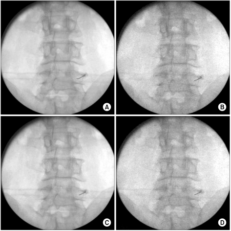
Fig. 4 According to C-arm modes, four fluoroscopic images blocking lumbar medial branch nerves. (A) Convention mode. (B) Pulsed mode. (C) Low-dose mode. (D) Pulsed + low-dose mode.
Cited by 9 articles
-
Radiation Safety for Pain Physicians
Sang Wook Shin
Korean J Pain. 2012;25(2):130-130. doi: 10.3344/kjp.2012.25.2.130.The Survey about the Degree of Damage of Radiation-Protective Shields in Operation Room
Jae Sung Ryu, Seung Woo Baek, Cheol Hee Jung, Suk Ju Cho, Eu Gene Jung, Hae Kyoung Kim, Jae Hun Kim
Korean J Pain. 2013;26(2):142-147. doi: 10.3344/kjp.2013.26.2.142.A Randomized Controlled Trial about the Levels of Radiation Exposure Depends on the Use of Collimation C-arm Fluoroscopic-guided Medial Branch Block
Seung Woo Baek, Jae Sung Ryu, Cheol Hee Jung, Joo Han Lee, Won Kyoung Kwon, Nam Sik Woo, Hae Kyoung Kim, Jae Hun Kim
Korean J Pain. 2013;26(2):148-153. doi: 10.3344/kjp.2013.26.2.148.Radiation Exposure of the Hand and Chest during C-arm Fluoroscopy-Guided Procedures
Cheol Hee Jung, Jae Sung Ryu, Seung Woo Baek, Ji Hye Oh, Nam Sik Woo, Hae Kyoung Kim, Jae Hun Kim
Korean J Pain. 2013;26(1):51-56. doi: 10.3344/kjp.2013.26.1.51.Radiation Safety for Pain Physicians: Technique or Equipment
Jae Hang Shim
Korean J Pain. 2014;27(2):101-102. doi: 10.3344/kjp.2014.27.2.101.The Radiation Exposure of Radiographer Related to the Location in C-arm Fluoroscopy-guided Pain Interventions
Young Jae Chang, Ah Na Kim, In Su Oh, Nam Sik Woo, Hae Kyoung Kim, Jae Hun Kim
Korean J Pain. 2014;27(2):162-167. doi: 10.3344/kjp.2014.27.2.162.How Effective Are Radiation Reducing Gloves in C-arm Fluoroscopy-guided Pain Interventions?
Ah Na Kim, Young Jae Chang, Bo Kyung Cheon, Jae Hun Kim
Korean J Pain. 2014;27(2):145-151. doi: 10.3344/kjp.2014.27.2.145.The radiation safety education and the pain physicians' efforts to reduce radiation exposure
Tae Hee Kim, Seung Wan Hong, Nam Sik Woo, Hae Kyoung Kim, Jae Hun Kim
Korean J Pain. 2017;30(2):104-115. doi: 10.3344/kjp.2017.30.2.104.Radiation safety for pain physicians: principles and recommendations
Sewon Park, Minjung Kim, Jae Hun Kim
Korean J Pain. 2022;35(2):129-139. doi: 10.3344/kjp.2022.35.2.129.
Reference
-
1. Raj PP, Lou L, Erdine S, Staats PS, Waldman SD, Racz G, et al. Interventional pain management: Image-guided procedures. 2008. 2nd ed. Philadelphia: Saunders Elsevier;p. 11.2. Kim TW, Jung JH, Jeon HJ, Yoon KB, Yoon DM. Radiation exposure to physicians during interventional pain procedures. Korean J Pain. 2010; 23:24–27. PMID: 20552069.
Article3. Goodman BS, Carnel CT, Mallempati S, Agarwal P. Reduction in average fluoroscopic exposure times for interventional spinal procedures through the use of pulsed and low-dose image settings. Am J Phys Med Rehabil. 2011; [Epub ahead of print].
Article4. Cousins C, Sharp C. Medical interventional procedures-reducing the radiation risks. Clin Radiol. 2004; 59:468–473. PMID: 15145716.
Article5. Mroz TE, Yamashita T, Davros WJ, Lieberman IH. Radiation exposure to the surgeon and the patient during kyphoplasty. J Spinal Disord Tech. 2008; 21:96–100. PMID: 18391712.
Article6. Miller DL, Vañó E, Bartal G, Balter S, Dixon R, Padovani R, et al. Occupational radiation protection in interventional radiology: a joint guideline of the Cardiovascular and Interventional Radiology Society of Europe and the Society of Interventional Radiology. Cardiovasc Intervent Radiol. 2010; 33:230–239. PMID: 20020300.
Article7. Brown PH, Thomas RD, Silberberg PJ, Johnson LM. Optimization of a fluoroscope to reduce radiation exposure in pediatric imaging. Pediatr Radiol. 2000; 30:229–235. PMID: 10789900.
Article8. Cohen MD. Can we use pulsed fluoroscopy to decrease the radiation dose during video fluoroscopic feeding studies in children? Clin Radiol. 2009; 64:70–73. PMID: 19070700.
Article9. Cohen MD. Optimizing the use of pulsed fluoroscopy to reduce radiation exposure to children. J Am Coll Radiol. 2008; 5:205–209. PMID: 18312969.
Article10. Rathmell JP. Atlas of image-guided intervention in regional anesthesia and pain medicine. 2006. Philadelphia: Lippincott Williams & Wilkins;p. 9–15.11. Uppot RN, Sahani DV, Hahn PF, Kalra MK, Saini SS, Mueller PR. Effect of obesity on image quality: fifteen-year longitudinal study for evaluation of dictated radiology reports. Radiology. 2006; 240:435–439. PMID: 16801372.
Article12. Wagner LK, Archer BR, Cohen AM. Management of patient skin dose in fluoroscopically guided interventional procedures. J Vasc Interv Radiol. 2000; 11:25–33. PMID: 10693710.
Article
- Full Text Links
-
- Actions
-
Cited
- CITED
-
- Close
- Share
-
- Similar articles
-
- A Randomized Controlled Trial about the Levels of Radiation Exposure Depends on the Use of Collimation C-arm Fluoroscopic-guided Medial Branch Block
- Radiation absorbed doses of cone beam computed tomography
- Estimating the Absorbed Dose to Critical Organs During Dual X-ray Absorptiometry
- Radiation exposure and its reduction in the fluoroscopic examination and fluoroscopy-guided interventional radiology
- Fluoroscopic Radiation Exposure during Percutaneous Kyphoplasty
