Comparison between Ultrasonography and Computed Tomography for Detecting the Pyramidal Lobe of the Thyroid Gland: A Prospective Multicenter Study
- Affiliations
-
- 1Department of Radiology, Busan Paik Hospital, Inje University College of Medicine, Busan 614-735, Korea.
- 2Department of Radiology, Seoul St. Mary's Hospital, College of Medicine, The Catholic University of Korea, Seoul 137-701, Korea. sljung1@catholic.ac.kr
- 3Department of Radiology, Severance Hospital, Yonsei University College of Medicine, Seoul 120-752, Korea.
- 4Department of Radiology, Haeundae Paik Hospital, Inje University College of Medicine, Busan 612-896, Korea.
- 5Department of Radiology, Thyroid Center, Daerim St. Mary's Hospital, Seoul 150-822, Korea.
- 6Department of Radiology, Soonchunhyang University Seoul Hospital, Soonchunhyang University College of Medicine, Seoul 140-743, Korea.
- KMID: 2070184
- DOI: http://doi.org/10.3348/kjr.2015.16.2.402
Abstract
OBJECTIVE
To compare the detection rates of the pyramidal lobe of the thyroid gland (TPL) using ultrasonography (US) and computed tomography (CT) in a prospective multi-center study.
MATERIALS AND METHODS
We enrolled 582 patients who underwent neck CT at six institutions. Each radiologist prospectively evaluated the presence and features of TPLs on thyroid US. Radiologists were divided into two groups according to their previous experience in detecting TPL on US or CT. The same radiologist also retrospectively assessed CT findings, blinded to the corresponding US findings.
RESULTS
The pyramidal lobe of the thyroid glands were detected in 230 cases (39.5%) on US and in 276 cases (47.6%) on CT. The TPL detection rate at the six institutions ranged from 22.0% to 59% for US and from 34.1% to 59% using CT. There were significant differences between US and CT in the detection rate, length, anteroposterior diameter, volume, and superior extent of TPL (p < or = 0.027). The TPL detection rates on both US and CT (p < 0.001) differed significantly according to the experience level of the radiologists. When the CT result was used as a reference standard, the sensitivity, specificity, positive and negative predictive values, as well as the accuracy of US for TPL detection were 72.6%, 91.5%, 89.3%, 77.3%, and 82.1%, respectively.
CONCLUSION
Our prospective multicenter study revealed that US could detect TPL with relatively high diagnostic accuracy compared to CT. Because the detection rate of TPL varied significantly according to the radiologists' level of experience, careful inspection is necessary to avoid imaging pitfalls and ensure appropriate evaluation of TPL on both US and CT.
Keyword
MeSH Terms
Figure
-

Fig. 1 Left pyramidal lobe detected on US and CT in 58-year-old woman. Transverse (A) and longitudinal (B) gray-scale sonograms show left pyramidal lobe (arrows). Non-enhanced (C) and contrast-enhanced (D) axial CT images of left pyramidal lobe (arrows) show same attenuation and enhancement as main thyroid gland (arrowheads).
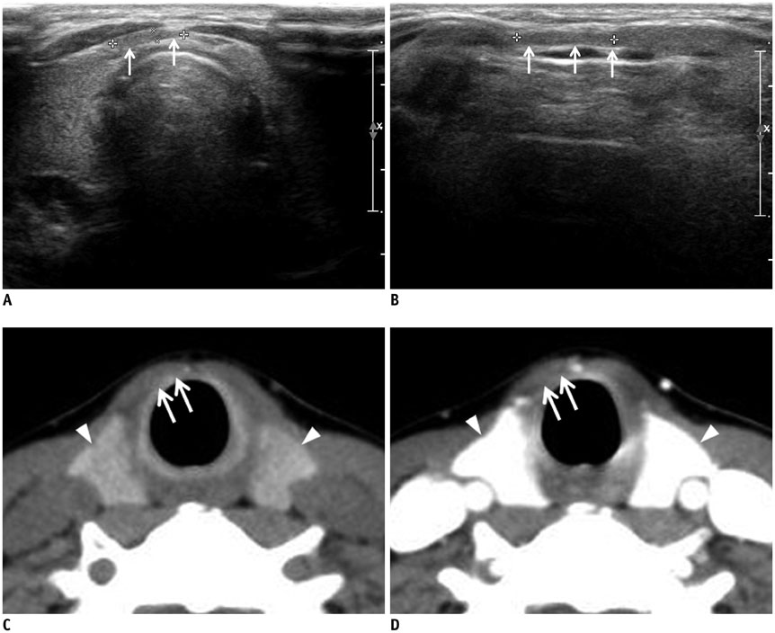
Fig. 2 Fatty tissue mimicking thyroid pyramidal lobe on ultrasonography in 49-year-old woman. Transverse (A) and longitudinal (B) gray-scale sonograms show longitudinally arranged structure mimicking thyroid pyramidal lobe (arrows). Nonenhanced (C) and contrast-enhanced (D) axial CT images shows only fatty tissue in same position (arrows), and they show different attenuation and enhancement from main thyroid gland (arrowheads).
Reference
-
1. Braun EM, Windisch G, Wolf G, Hausleitner L, Anderhuber F. The pyramidal lobe: clinical anatomy and its importance in thyroid surgery. Surg Radiol Anat. 2007; 29:21–27.2. Sultana S, Mannan S, Ahmed M, Rahman M, Khan M, Khalil M. An anatomical study on pyramidal lobe of thyroid gland in Bangladeshi people. Mymensingh Med J. 2008; 17:8–13.3. Ranade AV, Rai R, Pai MM, Nayak SR, Prakash , Krisnamurthy A, et al. Anatomical variations of the thyroid gland: possible surgical implications. Singapore Med J. 2008; 49:831–834.4. Geraci G, Pisello F, Li Volsi F, Modica G, Sciumè C. The importance of pyramidal lobe in thyroid surgery. G Chir. 2008; 29:479–482.5. Joshi SD, Joshi SS, Daimi SR, Athavale SA. The thyroid gland and its variations: a cadaveric study. Folia Morphol (Warsz). 2010; 69:47–45.6. Zivic R, Radovanovic D, Vekic B, Markovic I, Dzodic R, Zivaljevic V. Surgical anatomy of the pyramidal lobe and its significance in thyroid surgery. S Afr J Surg. 2011; 49:110.7. Ozgur Z, Celik S, Govsa F, Ozgur T. Anatomical and surgical aspects of the lobes of the thyroid glands. Eur Arch Otorhinolaryngol. 2011; 268:1357–1363.8. Pacini F, Schlumberger M, Harmer C, Berg GG, Cohen O, Duntas L, et al. Post-surgical use of radioiodine (131I) in patients with papillary and follicular thyroid cancer and the issue of remnant ablation: a consensus report. Eur J Endocrinol. 2005; 153:651–659.9. Park JY, Kim DW, Park JS, Kang T, Kim YW. The prevalence and features of thyroid pyramidal lobes as assessed by computed tomography. Thyroid. 2012; 22:173–177.10. Kim DW, Jung SL, Baek JH, Kim J, Ryu JH, Na DG, et al. The prevalence and features of thyroid pyramidal lobe, accessory thyroid, and ectopic thyroid as assessed by computed tomography: a multicenter study. Thyroid. 2013; 23:84–91.11. Kim KS, Kim DW, Sung JY. Detection of thyroid pyramidal lobe by ultrasound versus computed tomography: a single-center study. J Comput Assist Tomogr. 2014; 38:464–468.12. Kim DW, Ha TK, Park HK, Kang T. Sonographic detection of thyroid pyramidal lobes before thyroid surgery: a prospective single-center study. J Ultrasound Med. 2014; 33:239–244.13. Ryu JH, Kim DW, Kang T. Pre-operative detection of thyroid pyramidal lobes by ultrasound and computed tomography. Ultrasound Med Biol. 2014; 40:1442–1446.14. Torlontano M, Attard M, Crocetti U, Tumino S, Bruno R, Costante G, et al. Follow-up of low risk patients with papillary thyroid cancer: role of neck ultrasonography in detecting lymph node metastases. J Clin Endocrinol Metab. 2004; 89:3402–3407.15. Iida Y, Konishi J, Harioka T, Misaki T, Endo K, Torizuka K. Thyroid CT number and its relationship to iodine concentration. Radiology. 1983; 147:793–795.16. Frates MC, Benson CB, Charboneau JW, Cibas ES, Clark OH, Coleman BG, et al. Management of thyroid nodules detected at US: Society of Radiologists in Ultrasound consensus conference statement. Radiology. 2005; 237:794–800.17. Moon WJ, Baek JH, Jung SL, Kim DW, Kim EK, Kim JY, et al. Ultrasonography and the ultrasound-based management of thyroid nodules: consensus statement and recommendations. Korean J Radiol. 2011; 12:1–14.18. Ogawa C, Kammori M, Onose H, Yamada E, Shimizu K, Yamada T. Follicular carcinoma arising from the pyramidal lobe of the thyroid. J Nippon Med Sch. 2009; 76:169–172.
- Full Text Links
-
- Actions
-
Cited
- CITED
-
- Close
- Share
-
- Similar articles
-
- Papillary Carcinoma Arising from the Pyramidal Lobe of the Thyroid
- Papillary thyroid microcarcinoma in a thyroid pyramidal lobe
- Anatomical variations and developmental anomalies of the thyroid gland in Ethiopian population: a cadaveric study
- Morphologic Variations of the Thyroid Gland in Korean Adults
- CT Detection of Thyroid Pyramidal Lobe in Preoperative Patients with Thyroid Tumors
