J Adv Prosthodont.
2011 Dec;3(4):196-203. 10.4047/jap.2011.3.4.196.
A comparison of marginal fit of glass infiltrated alumina copings fabricated using two different techniques and the effect of firing cycles over them
- Affiliations
-
- 1Department of Prosthodontics, Sinhgad Dental College and Hospital, Pune, India. hirasankar@yahoo.com
- 2Saraswati-Dhanwantari Dental College and Hospital, Parbhani, India.
- KMID: 2054754
- DOI: http://doi.org/10.4047/jap.2011.3.4.196
Abstract
- PURPOSE
This study evaluated marginal fit of glass infiltrated alumina cores fabricated using two techniques and their marginal stability after firing cycles of veneering porcelain.
MATERIALS AND METHODS
Fifteen standardized all-ceramic crowns were fabricated on a metal die using each technique: slip cast technique of VITA In-Ceram sprint Alumina (Group A as control) and plastic foil matrix technique of Turkom-Cera fused alumina core system (Group B). Copings were compared between groups and within groups at coping stage and after firing each layer of veneering porcelain. A device was used to standardize seating of copings on the metal die and positioning of the specimens under the microscope after each stage of fabrication. The specimens were not cemented and marginal gap was measured using an image analyzing software (Imagepro Express) on the photographs captured under an optical microscope. Two tailed unpaired 't test' was used to compare marginal gaps in two groups and one way ANOVA was used to analyze marginal distortion within each group at 95% confidence interval.
RESULTS
The marginal gap was smaller at the coping stage in group B (60 + 30 microm) than group A (81 + 21 microm) with statistical significance. After firing of veneering porcelain the difference was insignificant. At the final stage, both groups exhibited lower mean marginal gaps than at the initial coping stage with the difference of 11.75 microm for group A and 11.94 microm for group B, but it was statistically insignificant due to high value of standard deviation.
CONCLUSION
Within the limitations of this study, it was concluded that both techniques produced copings with comparable and acceptable marginal fit and marginal stability on firing veneering porcelain.
MeSH Terms
Figure
-
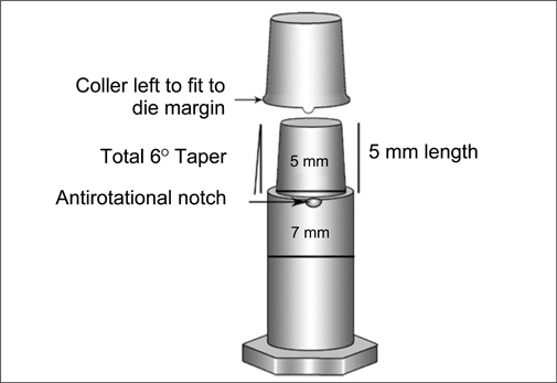
Fig. 1 Schematic diagram of metallic die and coping configurations.
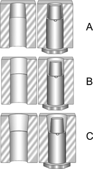
Fig. 2 Schematic diagram of metallic counter dies for porcelain buildup; A: base dentine, B: dentine and C: enamel.
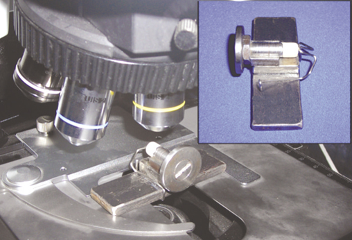
Fig. 3 Magnetic die orientation device and its placement under the microscope.
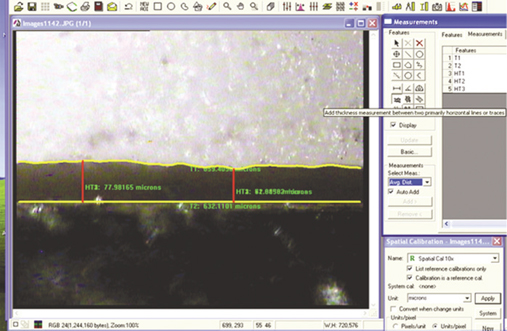
Fig. 4 Tracing of die and coping margins with image analyzing software.
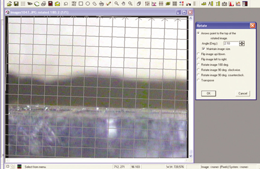
Fig. 5 Correction of rotated image with the image analyzer.
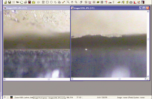
Fig. 6 Photographs captured at different focus level of same marginal area for superimposition.
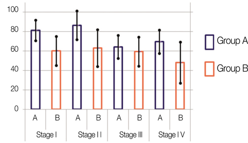
Fig. 7 Mean marginal gap in group A and B in four stages of fabrication.
Reference
-
1. Giordano RA. Dental ceramic restorative systems. Compend Contin Educ Dent. 1996. 17:779–782. 784–786. passim; quiz 794.2. Raigrodski AJ. Contemporary materials and technologies for all-ceramic fixed partial dentures: a review of the literature. J Prosthet Dent. 2004. 92:557–562.3. Conrad HJ, Seong WJ, Pesun IJ. Current ceramic materials and systems with clinical recommendations: a systematic review. J Prosthet Dent. 2007. 98:389–404.4. Felton DA, Kanoy BE, Bayne SC, Wirthman GP. Effect of in vivo crown margin discrepancies on periodontal health. J Prosthet Dent. 1991. 65:357–364.5. Sorensen JA. A rationale for comparison of plaque-retaining properties of crown systems. J Prosthet Dent. 1989. 62:264–269.6. Alkumru H, Hullah WR, Marquis PM, Wilson HJ. Factors affecting the fit of porcelain jacket crowns. Br Dent J. 1988. 164:39–43.7. Waerhaug J. Histologic considerations which govern where the margins of restorations should be located in relation to gingiva. Dent Clin North Am. 1960. 4:161–176.8. Löe H. Reactions to marginal periodontal tissues to restorative procedures. Int Dent J. 1968. 18:759–778.9. Orstavik D, Orstavik J. In vitro attachment of Streptococcus sanguis to dental crown and bridge cements. J Oral Rehabil. 1976. 3:139–144.10. Faucher RR, Nicholls JI. Distortion related to margin design in porcelain-fused-to-metal restorations. J Prosthet Dent. 1980. 43:149–155.11. Tuntiprawon M, Wilson PR. The effect of cement thickness on the fracture strength of all-ceramic crowns. Aust Dent J. 1995. 40:17–21.12. McLean JW, Hughes TH. The reinforcement of dental porcelain with ceramic oxides. Br Dent J. 1965. 119:251–267.13. Jones DW. Development of dental ceramics. An historical perspective. Dent Clin North Am. 1985. 29:621–644.14. Pera P, Gilodi S, Bassi F, Carossa S. In vitro marginal adaptation of alumina porcelain ceramic crowns. J Prosthet Dent. 1994. 72:585–590.15. Sorensen JA. A standardized method for determination of crown margin fidelity. J Prosthet Dent. 1990. 64:18–24.16. Lui JL. The effect of firing shrinkage on the marginal fit of porcelain jacket crowns. Br Dent J. 1980. 149:43–45.17. Sulaiman F, Chai J, Jameson LM, Wozniak WT. A comparison of the marginal fit of In-Ceram, IPS Empress, and Procera crowns. Int J Prosthodont. 1997. 10:478–484.18. Strating H, Pameijer CH, Gildenhuys RR. Evaluation of the marginal integrity of ceramometal restorations. Part I. J Prosthet Dent. 1981. 46:59–65.19. Christensen GJ. Marginal fit of gold inlay castings. J Prosthet Dent. 1966. 16:297–305.20. Shillingburg HT Jr, Hobo S, Fisher DW. Preparation design and margin distortion in porcelain-fused-to-metal restorations. J Prosthet Dent. 1973. 29:276–284.21. Buchanan WT, Svare CW, Turner KA. The effect of repeated firings and strength on marginal distortion in two ceramometal systems. J Prosthet Dent. 1981. 45:502–506.22. Denissen H, Dozić A, van der Zel J, van Waas M. Marginal fit and short-term clinical performance of porcelain-veneered CICERO, CEREC, and Procera onlays. J Prosthet Dent. 2000. 84:506–513.23. Yeo IS, Yang JH, Lee JB. In vitro marginal fit of three all-ceramic crown systems. J Prosthet Dent. 2003. 90:459–464.24. Att W, Komine F, Gerds T, Strub JR. Marginal adaptation of three different zirconium dioxide three-unit fixed dental prostheses. J Prosthet Dent. 2009. 101:239–247.25. Lofstrom LH, Barakat MM. Scanning electron microscopic evaluation of clinically cemented cast gold restorations. J Prosthet Dent. 1989. 61:664–669.26. Hamaguchi H, Cacciatore A, Tueller VM. Marginal distortion of the porcelain-bonded-to-metal complete crown: an SEM study. J Prosthet Dent. 1982. 47:146–153.27. Gemalmaz D, Alkumru HN. Marginal fit changes during porcelain firing cycles. J Prosthet Dent. 1995. 73:49–54.28. Balkaya MC, Cinar A, Pamuk S. Influence of firing cycles on the margin distortion of 3 all-ceramic crown systems. J Prosthet Dent. 2005. 93:346–355.29. May KB, Russell MM, Razzoog ME, Lang BR. Precision of fit: the Procera AllCeram crown. J Prosthet Dent. 1998. 80:394–404.30. McLean JW, von Fraunhofer JA. The estimation of cement film thickness by an in vivo technique. Br Dent J. 1971. 131:107–111.31. Chan C, Haraszthy G, Geis-Gerstorfer J, Weber H, Huettemann H. Scanning electron microscopic studies of the marginal fit of three esthetic crowns. Quintessence Int. 1989. 20:189–193.32. Vahidi F, Egloff ET, Panno FV. Evaluation of marginal adaptation of all-ceramic crowns and metal ceramic crowns. J Prosthet Dent. 1991. 66:426–431.33. Rinke S, Hüls A, Jahn L. Marginal accuracy and fracture strength of conventional and copy-milled all-ceramic crowns. Int J Prosthodont. 1995. 8:303–310.34. Hung SH, Hung KS, Eick JD, Chappell RP. Marginal fit of porcelain-fused-to-metal and two types of ceramic crown. J Prosthet Dent. 1990. 63:26–31.35. Davis DR. Comparison of fit of two types of all-ceramic crowns. J Prosthet Dent. 1988. 59:12–16.36. Weaver JD, Johnson GH, Bales DJ. Marginal adaptation of castable ceramic crowns. J Prosthet Dent. 1991. 66:747–753.37. Holmes JR, Sulik WD, Holland GA, Bayne SC. Marginal fit of castable ceramic crowns. J Prosthet Dent. 1992. 67:594–599.38. Shearer B, Gough MB, Setchell DJ. Influence of marginal configuration and porcelain addition on the fit of In-Ceram crowns. Biomaterials. 1996. 17:1891–1895.39. Boening KW, Wolf BH, Schmidt AE, Kästner K, Walter MH. Clinical fit of Procera AllCeram crowns. J Prosthet Dent. 2000. 84:419–424.40. Beschnidt SM, Strub JR. Evaluation of the marginal accuracy of different all-ceramic crown systems after simulation in the artificial mouth. J Oral Rehabil. 1999. 26:582–593.41. Chan C, Haraszthy G, Geis-Gerstofer J, Weber H. The marginal fit of Cerestore all ceramic crowns - A Primary report. Quintessence Int. 1985. 6:399–402.42. Komine F, Gerds T, Witkowski S, Strub JR. Influence of framework configuration on the marginal adaptation of zirconium dioxide ceramic anterior four-unit frameworks. Acta Odontol Scand. 2005. 63:361–366.43. Stappert CF, Dai M, Chitmongkolsuk S, Gerds T, Strub JR. Marginal adaptation of three-unit fixed partial dentures constructed from pressed ceramic systems. Br Dent J. 2004. 196:766–770. discussion 760quiz 78044. Grey NJ, Piddock V, Wilson MA. In vitro comparison of conventional crowns and a new all-ceramic system. J Dent. 1993. 21:47–51.45. Wolfart S, Wegner SM, Al-Halabi A, Kern M. Clinical evaluation of marginal fit of a new experimental all-ceramic system before and after cementation. Int J Prosthodont. 2003. 16:587–592.46. Reich S, Wichmann M, Nkenke E, Proeschel P. Clinical fit of all-ceramic three-unit fixed partial dentures, generated with three different CAD/CAM systems. Eur J Oral Sci. 2005. 113:174–179.47. Stappert CF, Dai M, Chitmongkolsuk S, Gerds T, Strub JR. Marginal adaptation of three-unit fixed partial dentures constructed from pressed ceramic systems. Br Dent J. 2004. 196:766–770. discussion 760quiz 78048. Ash MM, Nelson SJ. The Permanent Maxillary Premolars. Wheeler's Dental Anatomy, Physiology, and occlusion. 2003. 8th Ed. St. Louis: Sounder's (Imprint of Elsevier Pub.);243.49. Rosenstiel SF, Land MF, Fujimoto J. Contemporary Fixed Prosthodontics. 2002. 3rd Ed. St. Louis: Mosby (reprint), New Delhi Harcourt Private Limited;643–672.50. Holmes JR, Bayne SC, Holland GA, Sulik WD. Considerations in measurement of marginal fit. J Prosthet Dent. 1989. 62:405–408.51. Groten M, Axmann D, Pröbster L, Weber H. Determination of the minimum number of marginal gap measurements required for practical in-vitro testing. J Prosthet Dent. 2000. 83:40–49.
- Full Text Links
-
- Actions
-
Cited
- CITED
-
- Close
- Share
-
- Similar articles
-
- MARGINAL FIT OF GLASS INFILTRATED ALUMINA CORE FABRICATED FROM ALUMINA TAPES
- A STUDY ON THE DISTORTION OF THE COPINGS FOR CERAMOMETAL CROWNS DURING REPEATED FIRING
- Marginal fit related to margin types of glass infiltrated alumina core fabricated from aqueous-based alumina tape
- COMPARISON OF COLOR AND OPACITY OF COPY-MILLED IN-CREAM ALUMINA CORE AND SPINELL CORE
- FRACTURE STRENGTH AND MARGINAL FIT OF IN-CERAM, COPY-MILLED IN-CERAM, AND IPS EMPRESS 2 ALL-CERAMIC BRIDGES
