Imaging Sci Dent.
2015 Jun;45(2):81-87. 10.5624/isd.2015.45.2.81.
The effects of noise reduction, sharpening, enhancement, and image magnification on diagnostic accuracy of a photostimulable phosphor system in the detection of non-cavitated approximal dental caries
- Affiliations
-
- 1Department of Maxillofacial Radiology, Faculty of Dentistry, Guilan University of Medical Sciences, Rasht, Iran. zahradalili@yahoo.com
- 2Department of Operative Dentistry, Faculty of Dentistry, Guilan University of Medical Sciences, Rasht, Iran.
- 3Faculty of Dentistry, Guilan University of Medical Sciences, Rasht, Iran.
- KMID: 2054209
- DOI: http://doi.org/10.5624/isd.2015.45.2.81
Abstract
- PURPOSE
Contrast, sharpness, enhancement, and density can be changed in digital systems. The important question is to what extent the changes in these variables affect the accuracy of caries detection.
MATERIALS AND METHODS
Forty eight extracted human posterior teeth with healthy or proximal caries surfaces were imaged using a photostimulable phosphor (PSP) sensor. All original images were processed using a six-step method: (1) applying "Sharpening 2" and "Noise Reduction" processing options to the original images; (2) applying the "Magnification 1:3" option to the image obtained in the first step; (3) enhancing the original images by using the "Diagonal/" option; (4) reviewing the changes brought about by the third step of image processing and then, applying "Magnification 1:3"; (5) applying "Sharpening UM" to the original images; and (6) analyzing the changes brought about by the fifth step of image processing, and finally, applying "Magnification 1:3." Three observers evaluated the images. The tooth sections were evaluated histologically as the gold standard. The diagnostic accuracy of the observers was compared using a chi-squared test.
RESULTS
The accuracy levels irrespective of the image processing method ranged from weak (18.8%) to intermediate (54.2%), but the highest accuracy was achieved at the sixth image processing step. The overall diagnostic accuracy level showed a statistically significant difference (p=0.0001).
CONCLUSION
This study shows that the application of "Sharpening UM" along with the "Magnification 1:3" processing option improved the diagnostic accuracy and the observer agreement more effectively than the other processing procedures.
Figure
-
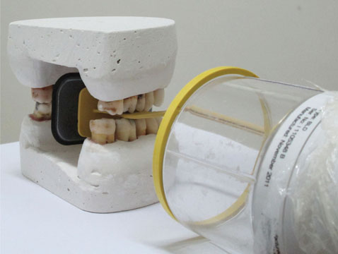
Fig. 1 Photograph shows the film positioning and tube setting method.

Fig. 2 Selection of the "Sharpening 2" option for the original image (A), and then, application of the "Noise Reduction" feature once (B).

Fig. 3 Enhancement of the original image by selecting the "Diagonal/" option (A), and then, application of "Magnification 1:3" (B).
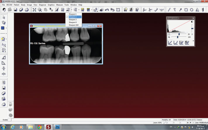
Fig. 4 Selection of the "Sharpening 2" option.
Reference
-
1. White SC, Pharoah MJ. Oral radiology; principles and interpretation. 6th ed. St Louis: Mosby;2014. p. 41.2. Wenzel A, Borg E, Hintze H, Gröndahl HG. Accuracy of caries diagnosis in digital images from charge-coupled device and storage phosphor systems: an in-vitro study. Dentomaxillofac Radiol. 1995; 24:250–254.3. Svanaes DB, Møystad A, Risnes S, Larheim TA, Gröndahl HG. Intraoral storage phosphor radiography for approximal caries detection and effect of image magnification: comparison with conventional radiography. Oral Surg Oral Med Oral Pathol Oral Radiol Endod. 1996; 82:94–100.4. Møystad A, Svanaes DB, Risnes S, Larheim TA, Gröndahl HG. Detection of approximal caries with a storage phosphor system. A comparison of enhanced digital images with dental X-ray film. Dentomaxillofac Radiol. 1996; 25:202–206.
Article5. Shrout MK, Russell CM, Potter BJ, Powell BJ, Hildebolt CF. Digital enhancement of radiographs: can it improve caries diagnosis? J Am Dent Assoc. 1996; 127:469–473.
Article6. Svanaes DB, Moystad A, Larheim TA. Approximal caries depth assessment with storage phosphor versus film radiography. Evaluation of the caries-specific Oslo enhancement procedure. Caries Res. 2000; 34:448–453.7. Li G, Yoshiura K, Welander U, Shi XQ, McDavid WD. Detection of approximal caries in digital radiographs before and after correction for attenuation and visual response. An in vitro study. Dentomaxillofac Radiol. 2002; 31:113–116.
Article8. Haak R, Wicht MJ, Nowak G, Hellmich M. Influence of displayed image size on radiographic detection of approximal caries. Dentomaxillofac Radiol. 2003; 32:242–246.
Article9. de Paola PF, Alman J. Assessment of the reliability of radiographic diagnosis in a clinical caries trial. J Dent Res. 1972; 51:1431–1437.
Article10. Crawley DA, Longbottom C, Cole BE, Ciesla CM, Arnone D, Wallace VP, et al. Terahertz pulse imaging: a pilot study of potential applications in dentistry. Caries Res. 2003; 37:352–359.
Article11. Bottenberg P, Jacquet W, Stachniss V, Wellnitz J, Schulte AG. Detection of cavitated or non-cavitated approximal enamel caries lesions using CMOS and CCD digital X-ray sensors and conventional D and F-speed films at different exposure conditions. Am J Dent. 2011; 24:74–78.12. Behere RR, Lele SM. Reliability of Logicon caries detector in the detection and depth assessment of dental caries: an in-vitro study. Indian J Dent Res. 2011; 22:362.
Article13. Syriopoulos K, Sanderink GC, Velders XL, van der Stelt PF. Radiographic detection of approximal caries: a comparison of dental films and digital imaging systems. Dentomaxillofac Radiol. 2000; 29:312–318.
Article14. Hintze H, Wenzel A, Larsen MJ. Stereomicroscopy, film radiography, microradiography and naked-eye inspection of tooth sections as validation for occlusal caries diagnosis. Caries Res. 1995; 29:359–363.
Article15. White SC, Pharoah MJ. Oral radiology; principles and interpretation. 6th ed. St Louis: Mosby;2014. p. 288.16. Abesi F, Mirshekar A, Moudi E, Seyedmajidi M, Haghanifar S, Haghighat N, et al. Diagnostic accuracy of digital and conventional radiography in the detection of non-cavitated approximal dental caries. Iran J Radiol. 2012; 9:17–21.
Article17. Seneadza V, Koob A, Kaltschmitt J, Staehle HJ, Duwenhoegger J, Eickholz P. Digital enhancement of radiographs for assessment of interproximal dental caries. Dentomaxillofac Radiol. 2008; 37:142–148.
Article18. Yoshiura K. Image quality assessment of digital intraoral radiography - perception to caries diagnosis. Jpn Dent Sci Rev. 2012; 48:42–47.
Article19. Naitoh M, Yuasa H, Toyama M, Shiojima M, Nakamura M, Ushida M, et al. Observer agreement in the detection of proximal caries with direct digital intraoral radiography. Oral Surg Oral Med Oral Pathol Oral Radiol Endod. 1998; 85:107–112.
Article
- Full Text Links
-
- Actions
-
Cited
- CITED
-
- Close
- Share
-
- Similar articles
-
- A Study on the Diagnostic Detection Ability of the Artificial Proximal Caries by Digora(R)
- Effect of changing the kilovoltage peak on radiographic caries assessment in digital and conventional radiography
- The influence of different scan resolutions on the detection of proximal caries lesions
- Effect of digital noise reduction on the accuracy of endodontic file length determination
- Image enhancement of digital periapical radiographs according to diagnostic tasks
