Minimum 1 Year Results of Arthroscopic Pull-out Repair for Posterior Root Tear of Medial Meniscus
- Affiliations
-
- 1Department of Orthopaedic Surgery, Daejeon Sun General Hospital, Daejeon, Korea. mydangjang@naver.com
- KMID: 2054001
- DOI: http://doi.org/10.5763/kjsm.2011.29.1.1
Abstract
- This study is to evaluate clinical and arthroscopic second-look results of arthroscopic repairs of posterior root tears of medial meniscus which may cause loss of circumferential hoop tension and extrusion of meniscus. From October 2006 to May 2009, fifty-eight patients (59 knees) underwent arthroscopic pull-out repairs. Clinical results were evaluated using Hospital for Special Surgery (HSS) score and International Knee Documentation Committee (IKDC) score for 12-month follow-up. Second-look arthroscopy was done to evaluate meniscal healing in 21 cases. Magnetic resonance imaging (MRI) was performed to assess status of repaired meniscus and tibial tunnel position in 9 patients. Average preoperative HSS score and IKDC score of 59 cases were 69.5 and 36.0, respectively. Average postoperative HSS score and IKDC score of 59 cases had been changed into 90.3 (p<0.001) and 66.8 (p<0.001), respectively. Second-look arthroscopies revealed complete or incomplete healing except one case. Two patients showed increased one grade according to the Kellgren-Lawrence radiologic classification system and others showed no change. Of 9 patients who performed MRI, six patients showed complete healing. The average position of tibial tunnel was 4.8 mm anterior and 5.7 mm medial to center of posterior cruciate ligament. Arthroscopic pull-out repair technique using transtibial tunnel seems to be simple and effective procedure for posterior root tear of medial meniscus. Further evaluation of arthroscopic repair of posterior root tear of medial meniscus should be needed to prove the effectiveness on the prevention of osteoarthritis of knee.
MeSH Terms
Figure
-
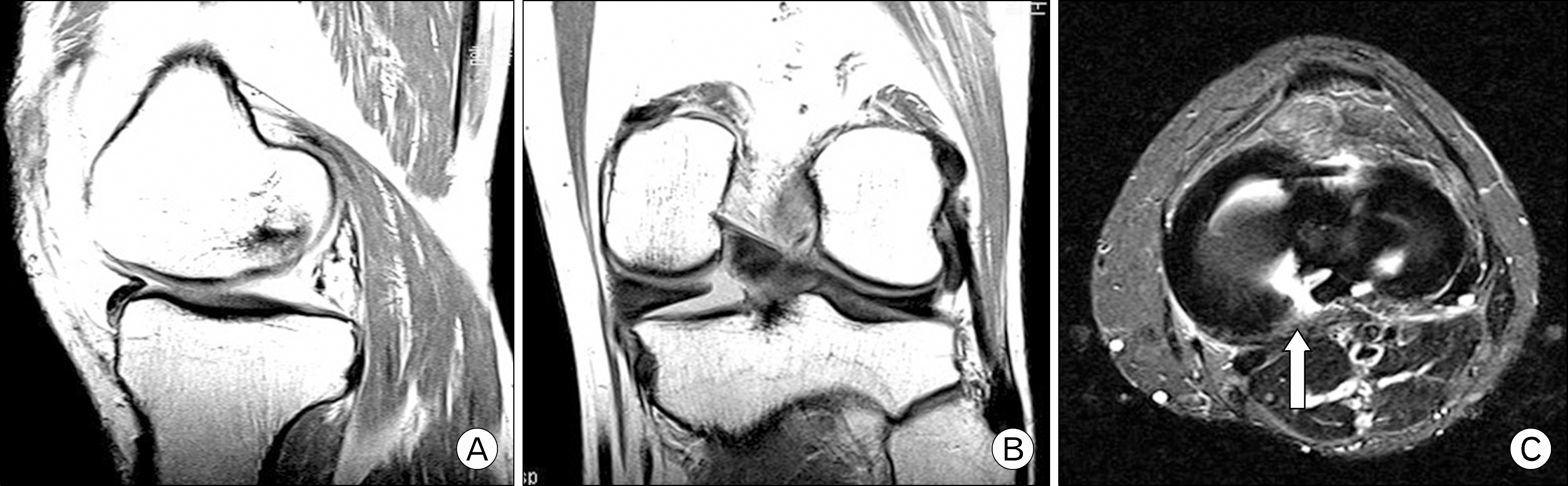
Fig. 1. Theses MRI findings demonstrated root tears of posterior horn of the medial meniscus on sagittal (ghost sign), coronal (cleft sign), and axial (cleavage sign, white arrow) section.
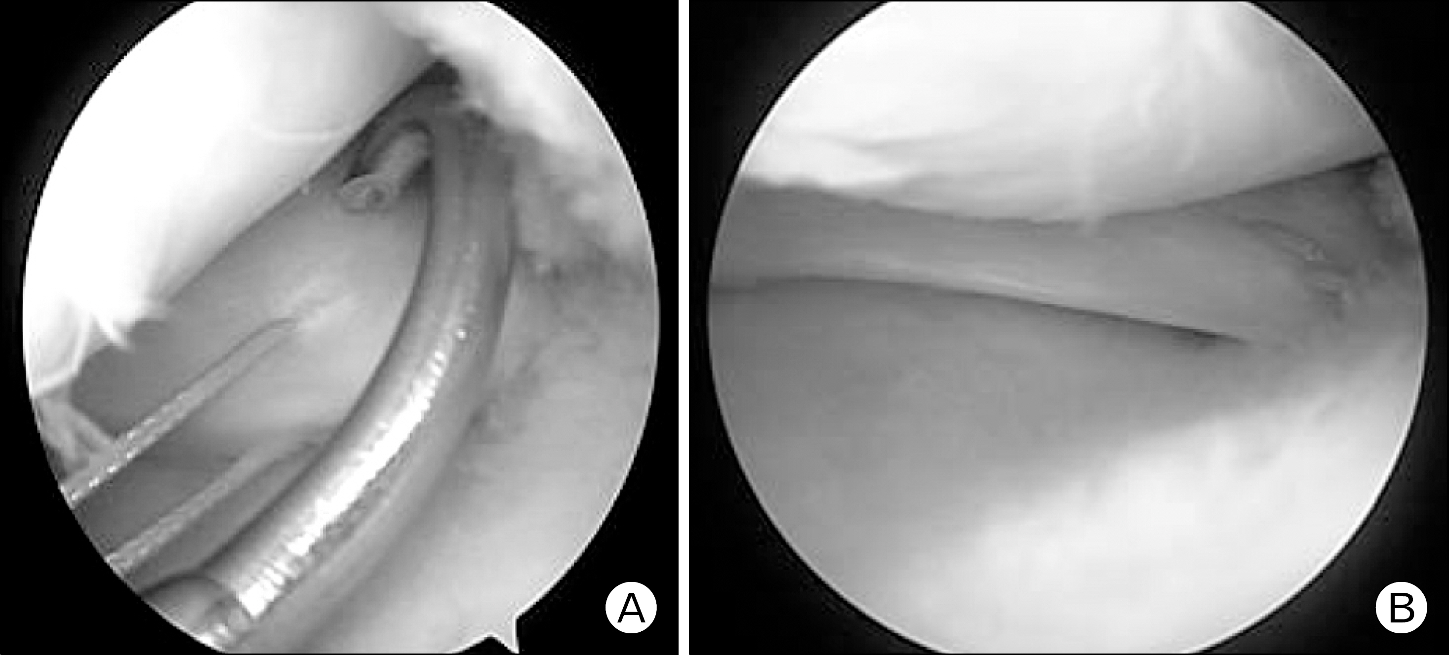
Fig. 2. (A) Polydioxanone No. 1 was advanced through the suture hook that penetrated the torn edge of the posterior horn of medial meniscus from supe-rior to inferior and changed to Ethibond No. 2 using shuttle relay technique. (B) Arthrosco-pic finding showed posterior horn of medial meniscus on the tibial foot print was well-re-attached.
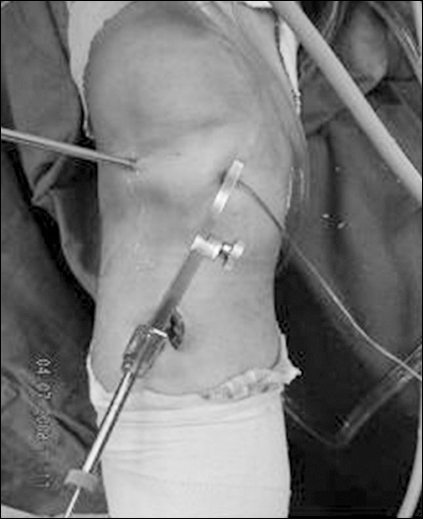
Fig. 3. The transosseous tibial tunnel was drilled from the anterolateral cortex of the proximal tibia to the insertion site of the posterior horn of medial meniscus using the anterior cruciate ligament tibial drilling guide.
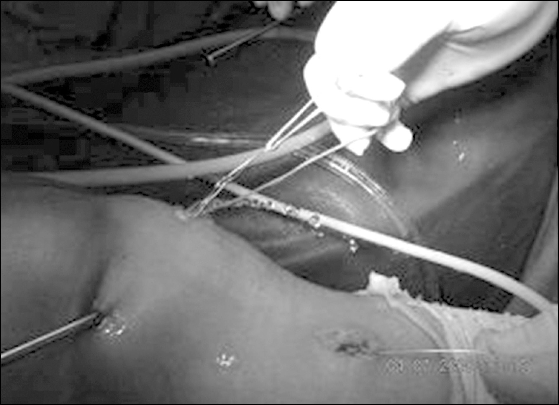
Fig. 4. All Ethibonds were brought out the tibial tunnel using advanced wire loop.
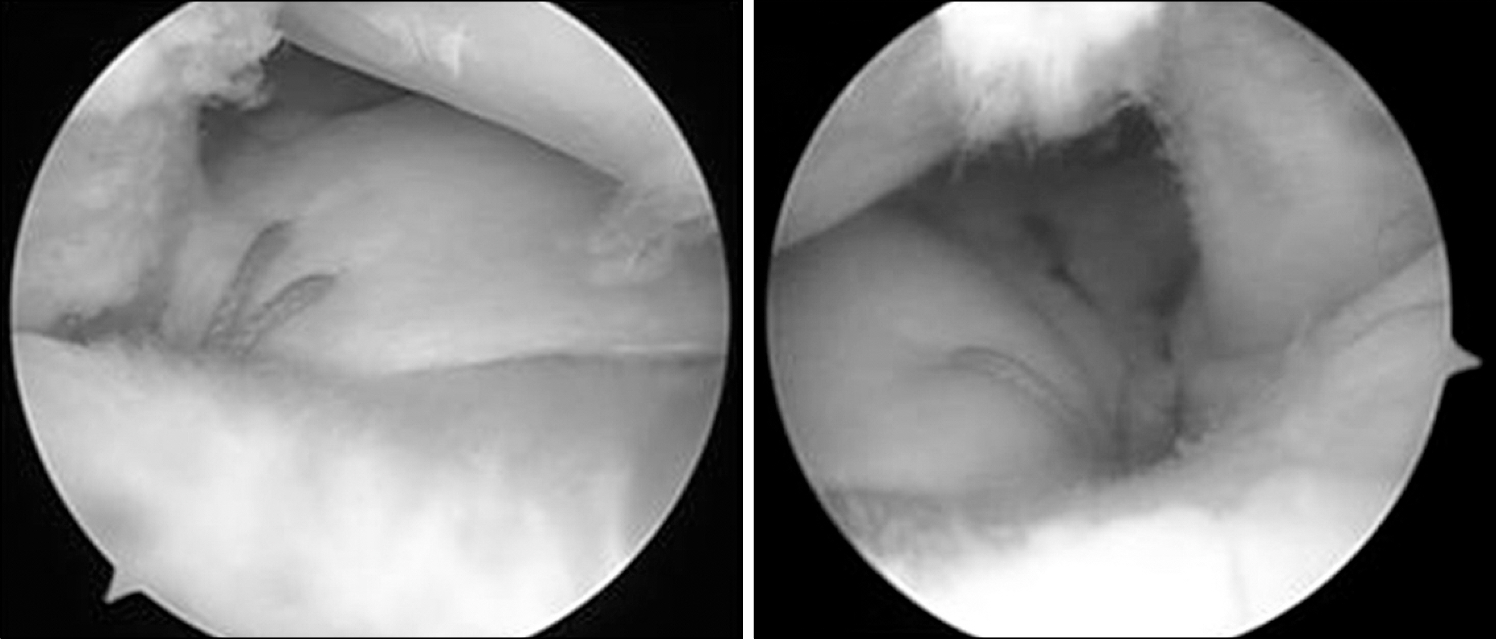
Fig. 5. Arthroscopy showed root of posterior horn of the me-niscus was reduced and well- reattached to the tibial insertion site.
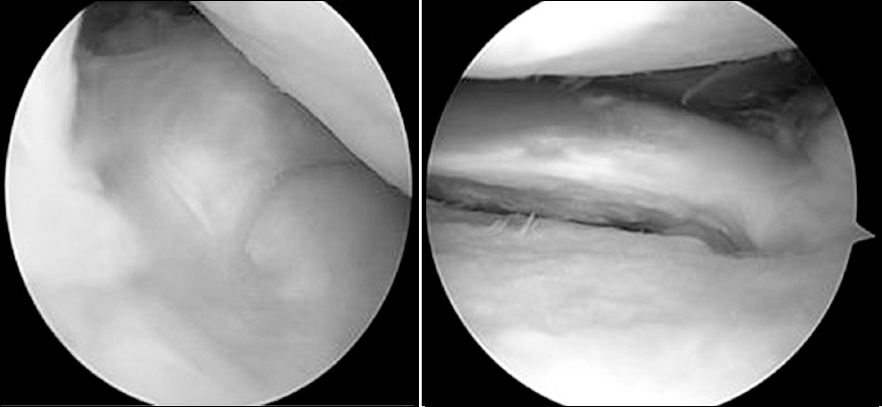
Fig. 6. Postoperative 6-month second-look arthroscopy re-vealed tear site was healed and reattached to the foot print of posterior horn of medial menis-cus.
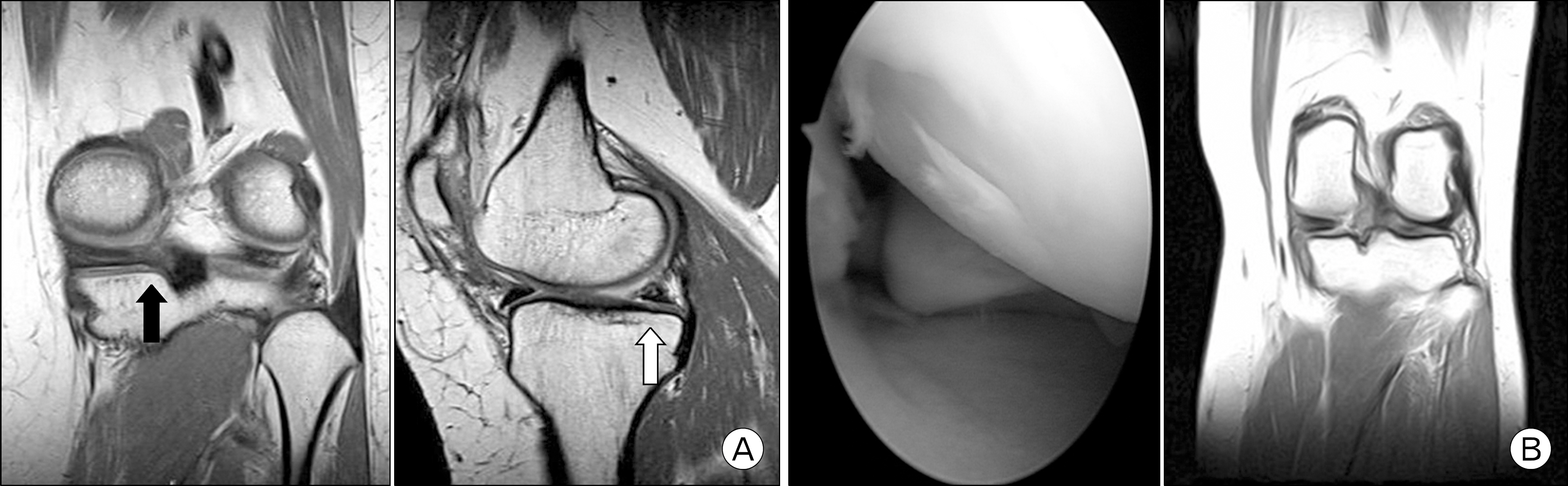
Fig. 7. (A) Postoperative MRI demonstrated intermediate signal density of healed meniscus tissue on foot print of posterior root of medial meniscus in coronal section (black arrow) and sagittal section (white arrow). (B) Second-look arthroscopy showed healing failure of medial meniscus and postoperative MRI demonstrated remained cleft sign on coronal section.

Fig. 8. Postoperative MRI showed entry point of transosseous tibial tunnel was located 4.8 mm anteriorly and 5.7 mm medially to center of posterior cruciate ligament insertion site. (A) axial section, (B) sagittal section, and (C) coronal section.
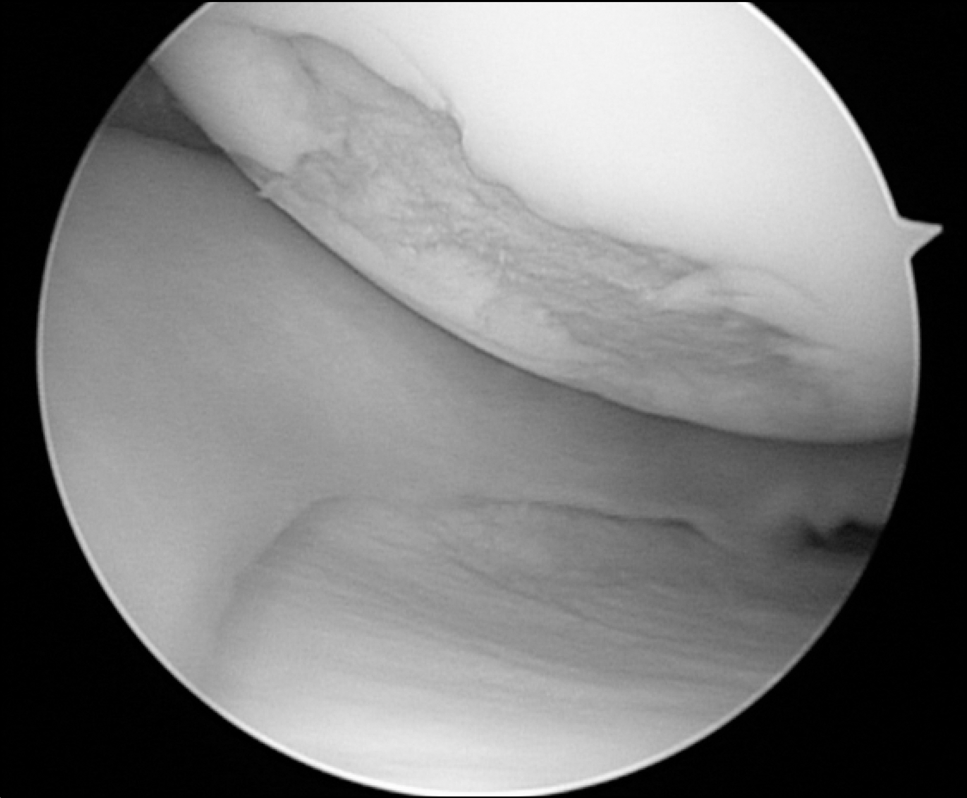
Fig. 9. Typical degenerative chondral lesion of medial femoral condyle due to root tear of posterior horn of me-dial meniscus showed irregular and moth-eaten appear-ance.
Reference
-
References
1. Ahn JH, Wang JH, Lim HC, et al. Double transosseous pull out suture technique for transection of posterior horn of medial meniscus. Arch Orthop Trauma Surg. 2009; 129:387–92.
Article2. Bessette GC. The meniscus. Orthopedics. 1992; 15:35–42.
Article3. Bin SI, Kim JM, Shin SJ. Radial tears of the posterior horn of the medial meniscus. Arthroscopy. 2004; 20:373–8.
Article4. Insall JN, Scott WN. Surgery of the knee. 3rd ed.New York: Churchill Livingstone;2001.5. Allaire R, Muriuki M, Gilbertson L, Harner CD. Biomechanical consequences of a tear of the posterior root of the medial meniscus. Similar to total meniscectomy. J Bone Joint Surg Am. 2008; 90:1922–31.6. Shino K, Hamada M, Mitsuoka T, Kinoshita H, Toritsuka Y. Arthroscopic repair for a flap tear of the posterior horn of the lateral meniscus adjacent to its tibial insertion. Arthroscopy. 1995; 11:495–8.
Article7. Ahn JH, Wang JH, Yoo JC, Noh HK, Park JH. A pull out suture for transection of the posterior horn of the medial meniscus: using a posterior trans-septal portal. Knee Surg Sports Traumatol Arthrosc. 2007; 15:1510–3.
Article8. Ozkoc G, Circi E, Gonc U, Irgit K, Pourbagher A, Tandogan RN. Radial tears in the root of the posterior horn of the medial meniscus. Knee Surg Sports Traumatol Arthrosc. 2008; 16:849–54.
Article9. Choi NH, Son KM, Victoroff BN. Arthroscopic all-inside repair for a tear of posterior root of the medial meniscus: a technical note. Knee Surg Sports Traumatol Arthrosc. 2008; 16:891–3.
Article10. Kim JG, Ha JG. Pullout repair of the radial posterior horn tear near the root of the medial meniscus: technical note. J Korean Knee Soc. 2004; 16:69–72.11. Kim DW, Moon JS, Kim MG, Kim JG. Pullout repair for root tear of medial meniscus. J Korean Arthrosc Soc. 2005; 9:40–5.12. Kim YM, Rhee KJ, Lee JK, Hwang DS, Yang JY, Kim SJ. Arthroscopic pullout repair of a complete radial tear of the tibial attachment site of the medial meniscus posterior horn. Arthroscopy. 2006; 22:795.e1–4.
Article13. Outerbridge RE. The etiology of chondromalacia patellae. J Bone Joint Surg Br. 1961; 43:752–7.
Article14. Kellgren JH, Lawrence JS. Radiological assessment of rheumatoid arthritis. Ann Rheum Dis. 1957; 16:485–93.
Article15. Nha KW, Jo JH, Lee DB. Clinical results of the radial tear of posterior root of medial meniscus. J Korean Arthrosc Soc. 2007; 11:128–33.16. Petersen W, Tillmann B. Age-related blood and lymph supply of the knee menisci. A cadaver study. Acta Orthop Scand. 1995; 66:308–12.17. Petersen W, Tillmann B. Collagenous fibril texture of the human knee joint menisci. Anat Embryol (Berl). 1998; 197:317–24.
Article18. Ahn JH, Kim SH, Yoo JC, Wang JH. All-inside suture technique using two posteromedial portals in a medial meniscus posterior horn tear. Arthroscopy. 2004; 20:101–8.
Article19. Wang YJ, Yu JK, Luo H, et al. An anatomical and histological study of human meniscal horn bony insertions and peri-meniscal attachments as a basis for meniscal transplantation. Chin Med J (Engl). 2009; 122:536–40.20. Lee JH, Lim YJ, Kim KB, Kim KH, Song JH. Arthroscopic pullout suture repair of posterior root tear of the medial meniscus: radiographic and clinical results with a 2-year follow-up. Arthroscopy. 2009; 25:951–8.
Article21. Cardello P, Gigli C, Ricci A, Chiatti L, Voglino N, Pofi E. Retears of postoperative knee meniscus: findings on magnetic resonance imaging (MRI) and magnetic resonance arthrography (MRA) by using low and high field magnets. Skeletal Radiol. 2009; 38:149–56.
Article22. Lee SY, Jee WH, Kim JM. Radial tear of the medial meniscal root: reliability and accuracy of MRI for diagnosis. AJR Am J Roentgenol. 2008; 191:81–5.
Article23. Marzo JM, Kumar BA. Primary repair of medial meniscal avulsions: 2 case studies. Am J Sports Med. 2007; 35:1380–3.
- Full Text Links
-
- Actions
-
Cited
- CITED
-
- Close
- Share
-
- Similar articles
-
- Pull-Out repair of the radial posterior horn tear near the root of the medial meniscus: Technical Note
- Non-Operative Treatment of the Degenerative Medial Meniscus Posterior Root Tear
- Modified Pull-out Suture in Posterior Root Tear of the Medial Meniscus: Using a Posteromedial Portal
- Arthroscopic Medial Meniscus Posterior Root Repair: Techniques and Current Issues
- Arthroscopic Direct Repair for a Complete Radial Tear of the Posterior Root of the Medial Meniscus
