Morphological assessment of the anterior loop of the mandibular canal in Koreans
- Affiliations
-
- 1Department of Anatomy and Orofacial Development, School of Dentistry, Chosun University, Gwangju, Korea. hjbkim@chosun.ac.kr
- KMID: 2046078
- DOI: http://doi.org/10.5115/acb.2015.48.1.75
Abstract
- The mandibular canal divides into the mental and incisive canals at the premolar region, forms the anterior loop which crosses anterior to the mental foramen, and turns back to reach the mental foramen. The aim of this study was to elucidate the general anatomical structure of the anterior loop of the mandibular canal using morphometry. Twenty-six hemimandibles from 19 cadavers (16 males, 3 females; mean age at death, 54.4 years) were studied by meticulous dissection with the aid of a surgical microscope. The location of the anterior loop, the diameters of the mandibular, mental, and incisive canals, and their distances from bony landmarks were measured using digital calipers. The anterior loop of the mandibular canal was located 3.05+/-1.15 mm (mean+/-SD) anterior to the anterior margin of the mental foramen and 2.72+/-1.41 mm inferior to the superior margin of the mental foramen, and was 4.34+/-1.46 mm long. The diameters of the mandibular, mental, and incisive canals were 2.8+/-0.49, 2.63+/-0.64, and 2.22+/-0.59 mm, respectively. The distances between the inferior border of the mandible and each of these canals were 7.82+/-1.52, 10.11+/-1.27, and 9.08+/-1.66 mm, respectively. The anterior loop of the mandibular canal was located a mean of 3.1 mm anterior and 2.7 mm inferior to the mental foramen, and continued upward and backward into the mental canal, and forward into the incisive canal. These detailed morphological features of the anterior loop of the mandibular canal represent useful practical anatomical knowledge regarding the interforaminal region.
Figure
-

Fig. 1 Photographs showing the origin definition of the anterior loop of mandibular canal. After determining the cutoff point of the neurovascular bundle, we devised a separated point of epineurium of each nerve as the anterior-most margin of anterior loop coincident with the origin of incisive canal. The inferior alveolar neurovascular bundle consists of the mental branch (black broken line) and incisive branch (white broken line).
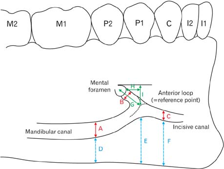
Fig. 2 Diagram showing the dimensions of the anterior loop measured. A, Diameter of the mandibular canal (10 mm back from the reference point); B, diameter of the mental canal; C, diameter of the incisive canal (5 mm forward of the reference point); D, distance from the mandibular inferior border to the inferior margin of the mandibular canal; E, distance from the mandibular inferior border to the inferior margin of the anterior loop; F, distance from the mandibular inferior border to the inferior margin of the incisive canal; G, length from the anterior loop to the mental foramen; H, horizontal distance from the anterior loop to the anterior margin of the mental foramen; I, vertical distance from the anterior loop to the superior margin of the mental foramen; I1, central incisor; I2, lateral incisor; C, canine; P1, first premolar; P2, second premolar; M1, first molar; M2, second molar.
Cited by 2 articles
-
A case of inferior alveolar nerve encircling the arteria maxillaris
Unnathi Nayak, Rajanigandha Vadgaonkar, Mangala M. Pai, B.V. Murlimanju
Anat Cell Biol. 2020;53(2):240-243. doi: 10.5115/acb.19.194.Assessment of the anterior loop of the inferior alveolar nerve via cone-beam computed tomography
Baratollah Shaban, Amin Khajavi, Nasim Khaki, Yones Mohiti, Tahere Mehri, Hamed Kermani
J Korean Assoc Oral Maxillofac Surg. 2017;43(6):395-400. doi: 10.5125/jkaoms.2017.43.6.395.
Reference
-
1. Bavitz JB, Harn SD, Hansen CA, Lang M. An anatomical study of mental neurovascular bundle-implant relationships. Int J Oral Maxillofac Implants. 1993; 8:563–567.2. Apostolakis D, Brown JE. The anterior loop of the inferior alveolar nerve: prevalence, measurement of its length and a recommendation for interforaminal implant installation based on cone beam CT imaging. Clin Oral Implants Res. 2012; 23:1022–1030.3. Juodzbalys G, Wang HL, Sabalys G. Anatomy of Mandibular Vital Structures. Part II: Mandibular Incisive Canal, Mental Foramen and Associated Neurovascular Bundles in Relation with Dental Implantology. J Oral Maxillofac Res. 2010; 1:e3.4. Drake RL, Vogl AW, Mitchell AWM. Gray's anatomy for students. 2nd ed. Philadelphia: Churchill Livingstone;2010. p. 1056–1060.5. Won SY, Kim DH, Yang HM, Park JT, Kwak HH, Hu KS, Kim HJ. Clinical and anatomical approach using Sihler's staining technique (whole mount nerve stain). Anat Cell Biol. 2011; 44:1–7.6. Mardinger O, Chaushu G, Arensburg B, Taicher S, Kaffe I. Anterior loop of the mental canal: an anatomical-radiologic study. Implant Dent. 2000; 9:120–125.7. Hwang K, Lee WJ, Song YB, Chung IH. Vulnerability of the inferior alveolar nerve and mental nerve during genioplasty: an anatomic study. J Craniofac Surg. 2005; 16:10–14.8. Greenstein G, Tarnow D. The mental foramen and nerve: clinical and anatomical factors related to dental implant placement: a literature review. J Periodontol. 2006; 77:1933–1943.9. Kim ST, Hu KS, Song WC, Kang MK, Park HD, Kim HJ. Location of the mandibular canal and the topography of its neurovascular structures. J Craniofac Surg. 2009; 20:936–939.10. Uchida Y, Yamashita Y, Goto M, Hanihara T. Measurement of anterior loop length for the mandibular canal and diameter of the mandibular incisive canal to avoid nerve damage when installing endosseous implants in the interforaminal region. J Oral Maxillofac Surg. 2007; 65:1772–1779.11. Li X, Jin ZK, Zhao H, Yang K, Duan JM, Wang WJ. The prevalence, length and position of the anterior loop of the inferior alveolar nerve in Chinese, assessed by spiral computed tomography. Surg Radiol Anat. 2013; 35:823–830.12. Hu KS, Yun HS, Hur MS, Kwon HJ, Abe S, Kim HJ. Branching patterns and intraosseous course of the mental nerve. J Oral Maxillofac Surg. 2007; 65:2288–2294.13. Watanabe H, Mohammad Abdul M, Kurabayashi T, Aoki H. Mandible size and morphology determined with CT on a premise of dental implant operation. Surg Radiol Anat. 2010; 32:343–349.14. Arzouman MJ, Otis L, Kipnis V, Levine D. Observations of the anterior loop of the inferior alveolar canal. Int J Oral Maxillofac Implants. 1993; 8:295–300.15. Kuzmanovic DV, Payne AG, Kieser JA, Dias GJ. Anterior loop of the mental nerve: a morphological and radiographic study. Clin Oral Implants Res. 2003; 14:464–471.16. Kaya Y, Sencimen M, Sahin S, Okcu KM, Dogan N, Bahcecitapar M. Retrospective radiographic evaluation of the anterior loop of the mental nerve: comparison between panoramic radiography and spiral computerized tomography. Int J Oral Maxillofac Implants. 2008; 23:919–925.17. Uchida Y, Noguchi N, Goto M, Yamashita Y, Hanihara T, Takamori H, Sato I, Kawai T, Yosue T. Measurement of anterior loop length for the mandibular canal and diameter of the mandibular incisive canal to avoid nerve damage when installing endosseous implants in the interforaminal region: a second attempt introducing cone beam computed tomography. J Oral Maxillofac Surg. 2009; 67:744–750.18. Chen JC, Lin LM, Geist JR, Chen JY, Chen CH, Chen YK. A retrospective comparison of the location and diameter of the inferior alveolar canal at the mental foramen and length of the anterior loop between American and Taiwanese cohorts using CBCT. Surg Radiol Anat. 2013; 35:11–18.19. Ngeow WC, Dionysius DD, Ishak H, Nambiar P. A radiographic study on the visualization of the anterior loop in dentate subjects of different age groups. J Oral Sci. 2009; 51:231–237.20. Jacobs R, Mraiwa N, Van Steenberghe D, Sanderink G, Quirynen M. Appearance of the mandibular incisive canal on panoramic radiographs. Surg Radiol Anat. 2004; 26:329–333.21. Mardinger O, Chaushu G, Arensburg B, Taicher S, Kaffe I. Anatomic and radiologic course of the mandibular incisive canal. Surg Radiol Anat. 2000; 22:157–161.22. Wical KE, Swoope CC. Studies of residual ridge resorption. I. Use of panoramic radiographs for evaluation and classification of mandibular resorption. J Prosthet Dent. 1974; 32:7–12.
- Full Text Links
-
- Actions
-
Cited
- CITED
-
- Close
- Share
-
- Similar articles
-
- Observation of the anterior loop and mental foramen of the mandibular canal using cone beam computed tomography
- Panoramic radiographs underestimate extensions of the anterior loop and mandibular incisive canal
- 3-dimensional reconstruction of mandibular canal at the interforaminal region using micro-computed tomography in Korean
- Observation of mandibular second molar roots and root canal morphology using dental cone-beam computed tomography
- Assessment of the anterior loop of the mandibular canal: A study using cone-beam computed tomography
