Bisphosphonate-related osteonecrosis of the jaw in a multiple myeloma patient: A case report with characteristic radiographic features
- Affiliations
-
- 1Department of Oral and Maxillofacial Radiology and Wonkwang Dental Research Institute, College of Dentistry, Wonkwang University, Iksan, Korea. eebydo@wonkwang.ac.kr
- 2Department of Internal Medicine, School of Medicine, Wonkwang University, Iksan, Korea.
- 3Department of Oral and Maxillofacial Surgery, College of Dentistry, Wonkwang University, Iksan, Korea.
- KMID: 2045029
- DOI: http://doi.org/10.5624/isd.2015.45.3.199
Abstract
- A 59-year-old male who had suffered from multiple myeloma for nine years and had been administered bisphosphonates for seven years visited a dental hospital for pain relief due to extensive caries in his left maxillary molars. The molars were extracted, leaving an exposed wound for three months. The radiograph showed sequestra formation and irregular bone destruction in the left maxilla. Sudden pain and gingival swelling in the right mandibular molar area occurred six months later. The interseptum of the right lower second molar was observed to be necrotic during surgery. These findings coincided with the features of bisphosphonate-related osteonecrosis of the jaw (BRONJ). In this case, the long intravenous administration of bisphosphonates and tooth extraction were likely the etiologic factors of BRONJ in a patient with multiple myeloma; moreover, the bilateral occurrence of BRONJ is a characteristic feature.
MeSH Terms
Figure
-
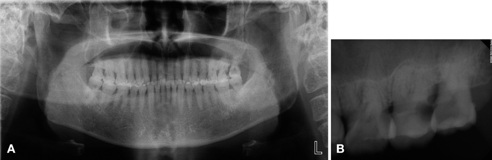
Fig. 1 A panoramic radiograph (A) and periapical radiograph (B) show a carious lesion on the maxillary first, second, and third molars extending into the subgingival area, with a suspected mucosal antral cyst in the left maxillary sinus.
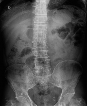
Fig. 2 Compression fracture of T12 and diffuse osteopenia of the pelvic bone.
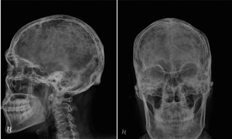
Fig. 3 Skull radiographs show a punched-out appearance.

Fig. 4 At 50 days after extraction, pus discharge from the wound and sequestra formation are observed.
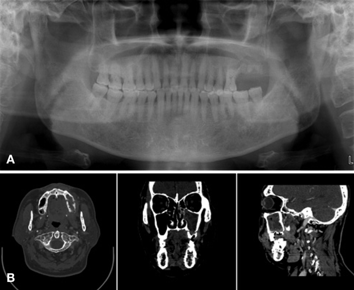
Fig. 5 A panoramic radiograph (A) and a computed tomograph (B) reveal the separation of the alveolar ridge from the surrounding maxillary bone, sequestra, and irregular bone destruction. Chronic sinusitis is seen in the left maxillary sinus.

Fig. 6 A photograph shows the extracted premolars, the first molar, and a necrotized bone fragment.

Fig. 7 A. An intraoral photograph shows gingival swelling around the lower second and third molars. B. A periapical radiograph shows moderate periodontitis and proximal caries on the distal surface of the first molar.
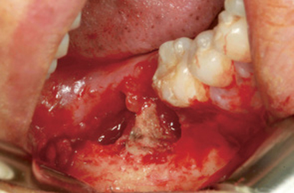
Fig. 8 A photograph shows the necrotic interseptum of the right lower second molar.
Cited by 1 articles
-
Clinical and panoramic radiographic features of osteomyelitis of the jaw: A comparison between antiresorptive medication-related and medication-unrelated conditions
Jeong Won Shin, Jo-Eun Kim, Kyung-Hoe Huh, Won-Jin Yi, Min-Suk Heo, Sam-Sun Lee, Soon-Chul Choi
Imaging Sci Dent. 2019;49(4):287-294. doi: 10.5624/isd.2019.49.4.287.
Reference
-
1. Kyle RA. Multiple myeloma: review of 869 cases. Mayo Clin Proc. 1975; 50:29–40.2. Batsakis JG. Pathology consultation. Plasma cell tumors of the head and neck. Ann Otol Rhinol Laryngol. 1983; 92:311–313.3. Wingo PA, Tong T, Bolden S. Cancer statistics, 1995. CA Cancer J Clin. 1995; 45:8–30.
Article4. Holland J, Trenkner DA, Wasserman TH, Fineberg B. Plasmacytoma. Treatment results and conversion to myeloma. Cancer. 1992; 69:1513–1517.
Article5. Kyle RA. Diagnostic criteria of multiple myeloma. Hematol Oncol Clin North Am. 1992; 6:347–358.
Article6. Lee SH, Huang JJ, Pan WL, Chan CP. Gingival mass as the primary manifestation of multiple myeloma: report of two cases. Oral Surg Oral Med Oral Pathol Oral Radiol Endod. 1996; 82:75–79.7. Epstein JB, Voss NJ, Stevenson-Moore P. Maxillofacial manifestations of multiple myeloma. An unusual case and review of the literature. Oral Surg Oral Med Oral Pathol. 1984; 57:267–271.8. Lambertenghi-Deliliers G, Bruno E, Cortelezzi A, Fumagalli L, Morosini A. Incidence of jaw lesions in 193 patients with multiple myeloma. Oral Surg Oral Med Oral Pathol. 1988; 65:533–537.9. Mondello P, Pitini V, Arrigo C, Mondello S, Mian M, Altavilla G. Necrotizing fasciitis as a rare complication of osteonecrosis of the jaw in a patient with multiple myeloma treated with lenalidomide: case report and review of the literature. Springerplus. 2014; 3:123.
Article10. Coleman R, Gnant M, Morgan G, Clezardin P. Effects of bone-targeted agents on cancer progression and mortality. J Natl Cancer Inst. 2012; 104:1059–1067.
Article11. Lee SH, Chan RC, Chang SS, Tan YL, Chang KH, Lee MC, et al. Use of bisphosphonates and the risk of osteonecrosis among cancer patients: a systemic review and meta-analysis of the observational studies. Support Care Cancer. 2014; 22:553–560.
Article12. Utreja A, Almas K, Javed F. Dental extraction as a risk factor for bisphosphonate related osteonecrosis of the jaw in cancer patients: an update. Odontostomatol Trop. 2013; 36:38–46.13. Sukpanichnant S, Cousar JB, Leelasiri A, Graber SE, Greer JP, Collins RD. Diagnostic criteria and histologic grading in multiple myeloma: histologic and immunohistologic analysis of 176 cases with clinical correlation. Hum Pathol. 1994; 25:308–318.
Article14. Bladé J, Kyle RA, Greipp PR. Multiple myeloma in patients younger than 30 years. Report of 10 cases and review of the literature. Arch Intern Med. 1996; 156:1463–1468.15. Matsumura S, Kishino M, Ishida T, Furukawa S. Radiographic findings for solitary plasmacytoma of the bone in the anterior wall of the maxillary sinus: a case report. Oral Surg Oral Med Oral Pathol Oral Radiol Endod. 2000; 89:651–657.
Article16. Croucher PI, Apperley JF. Bone disease in multiple myeloma. Br J Haematol. 1998; 103:902–910.
Article17. Mozaffari E, Mupparapu M, Otis L. Undiagnosed multiple myeloma causing extensive dental bleeding: report of a case and review. Oral Surg Oral Med Oral Pathol Oral Radiol Endod. 2002; 94:448–453.
Article18. Yaegaki K, Kameyama T, Takenaka M, Kimura T, Sujaku C, Tanimura A. Myelomatosis (IgD, lambda) discovered by oral manifestation. Int J Oral Surg. 1985; 14:381–384.19. Badros A, Weikel D, Salama A, Goloubeva O, Schneider A, Rapoport A. Osteonecrosis of the jaw in multiple myeloma patients: clinical features and risk factors. J Clin Oncol. 2006; 24:945–952.
Article20. Advisory Task Force on Bisphosphonate-Related Ostenonecrosis of the Jaws, American Association of Oral and Maxillofacial Surgeons. American Association of Oral and Maxillofacial Surgeons position paper on bisphosphonate-related osteonecrosis of the jaws. J Oral Maxillofac Surg. 2007; 65:369–376.21. Assael LA. New foundations in understanding osteonecrosis of the jaws. J Oral Maxillofac Surg. 2004; 62:125–126.
Article
- Full Text Links
-
- Actions
-
Cited
- CITED
-
- Close
- Share
-
- Similar articles
-
- A Case of Sinusitis due to Bisphosphonate Related Osteonecrosis of Jaw
- Clinical feature and treatment of bisphosphonate-related osteonecrosis of jaw about oral bisphosphonate administrated patients: case reports
- Multidisciplinary approach for medication-related osteonecrosis of the jaws: a case report and literature review
- A case of bisphosphonate-related osteonecrosis of the jaw with a particularly unfavourable course: a case report
- A Case of Intractable Bisphosphonate-Related Osteonecrosis of the Jaw Treated with Teriparatide
