Imaging Sci Dent.
2015 Sep;45(3):153-158. 10.5624/isd.2015.45.3.153.
Accuracy of digital periapical radiography and cone-beam computed tomography in detecting external root resorption
- Affiliations
-
- 1Division of Dental Diagnostic Science, Rutgers School of Dental Medicine, Newark, NJ, USA. adriana.creanga@rutgers.edu
- 2Division of Oral and Maxillofacial Radiology, Department of Comprehensive Dentistry, University of Texas Health Science Center San Antonio, San Antonio, TX, USA.
- 3Oral Medicine Clinic, University of Texas Health Science Center San Antonio, San Antonio, TX, USA.
- 4Department of Endodontics, University of Iowa, Iowa City, IA, USA.
- 5Department of Pathology, University of Texas Health Science Center San Antonio, San Antonio, TX, USA.
- KMID: 2045022
- DOI: http://doi.org/10.5624/isd.2015.45.3.153
Abstract
- PURPOSE
The purpose of this study was to evaluate and compare the efficacy of cone-beam computed tomography (CBCT) and digital intraoral radiography in diagnosing simulated small external root resorption cavities.
MATERIALS AND METHODS
Cavities were drilled in 159 roots using a small spherical bur at different root levels and on all surfaces. The teeth were imaged both with intraoral digital radiography using image plates and with CBCT. Two sets of intraoral images were acquired per tooth: orthogonal (PA) which was the conventional periapical radiograph and mesioangulated (SET). Four readers were asked to rate their confidence level in detecting and locating the lesions. Receiver operating characteristic (ROC) analysis was performed to assess the accuracy of each modality in detecting the presence of lesions, the affected surface, and the affected level. Analysis of variation was used to compare the results and kappa analysis was used to evaluate interobserver agreement.
RESULTS
A significant difference in the area under the ROC curves was found among the three modalities (P=0.0002), with CBCT (0.81) having a significantly higher value than PA (0.71) or SET (0.71). PA was slightly more accurate than SET, but the difference was not statistically significant. CBCT was also superior in locating the affected surface and level.
CONCLUSION
CBCT has already proven its superiority in detecting multiple dental conditions, and this study shows it to likewise be superior in detecting and locating incipient external root resorption.
MeSH Terms
Figure
-
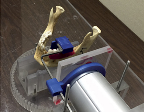
Fig. 1 A photograph shows the use of soft tissue simulation in acquiring intraoral radiographs.
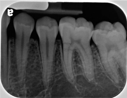
Fig. 2 Orthogonal periapical radiograph (PA) is seen.

Fig. 3 A set of orthogonal (PA, left) and corresponding mesioangulated projection (SET, right) are taken.
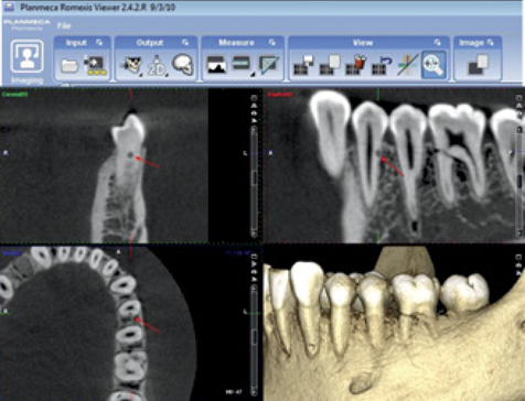
Fig. 4 Planmeca Romexis Viewer Software shows axial, coronal, sagittal, and three-dimensional CBCT images of simulated external root resorption (arrows).
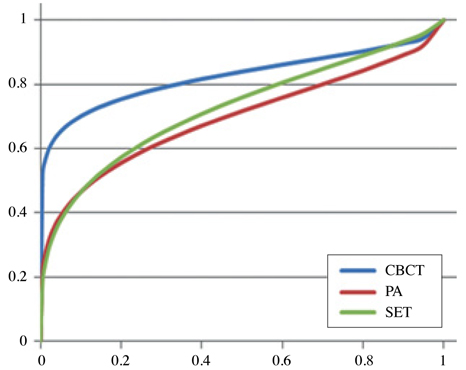
Fig. 5 Receiver operating characteristic (ROC) curves for all modalities of PA, SET, and CBCT images.
Reference
-
1. Fuss Z, Tsesis I, Lin S. Root resorption - diagnosis, classification and treatment choices based on stimulation factors. Dent Traumatol. 2003; 19:175–182.
Article2. Patel S, Dawood A, Wilson R, Horner K, Mannocci F. The detection and management of root resorption lesions using intraoral radiography and cone beam computed tomography - an in vivo investigation. Int Endod J. 2009; 42:831–838.3. Patel S, Dawood A, Ford TP, Whaites E. The potential applications of cone beam computed tomography in the management of endodontic problems. Int Endod J. 2007; 40:818–830.
Article4. Noujeim M, Prihoda T, Langlais R, Nummikoski P. Evaluation of high-resolution cone beam computed tomography in the detection of simulated interradicular bone lesions. Dentomaxillofac Radiol. 2009; 38:156–162.
Article5. Durack C, Patel S, Davies J, Wilson R, Mannocci F. Diagnostic accuracy of small volume cone beam computed tomography and intraoral periapical radiography for the detection of simulated external inflammatory root resorption. Int Endod J. 2011; 44:136–147.
Article6. Bernardes RA, de Paulo RS, Pereira LO, Duarte MA, Ordinola-Zapata R, de Azevedo JR. Comparative study of cone beam computed tomography and intraoral periapical radiographs in diagnosis of lingual-simulated external root resorptions. Dent Traumatol. 2012; 28:268–272.
Article7. Kamburoğlu K, Kurşun S, Yüksel S, Oztaş B. Observer ability to detect ex vivo simulated internal or external cervical root resorption. J Endod. 2011; 37:168–175.
Article8. D'Addazio PS, Campos CN, Özcan M, Teixeira HG, Passoni RM, Carvalho AC. A comparative study between cone-beam computed tomography and periapical radiographs in the diagnosis of simulated endodontic complications. Int Endod J. 2011; 44:218–224.9. Kumar V, Gossett L, Blattner A, Iwasaki LR, Williams K, Nickel JC. Comparison between cone-beam computed tomography and intraoral digital radiography for assessment of tooth root lesions. Am J Orthod Dentofacial Orthop. 2011; 139:e533–e541.
Article10. American Association of Endodontists. American Academy of Oral and Maxillofacial Radiography. AAE and AAOMR joint position statement. Use of cone-beam-computed tomography in endodontics. Pa Dent J (Harrisb). 2011; 78:37–39.11. Ludlow JB, Davies-Ludlow LE, White SC. Patient risk related to common dental radiographic examinations: the impact of 2007 International Commission on Radiological Protection recommendations regarding dose calculation. J Am Dent Assoc. 2008; 139:1237–1243.12. Qu XM, Li G, Ludlow JB, Zhang ZY, Ma XC. Effective radiation dose of ProMax 3D cone-beam computerized tomography scanner with different dental protocols. Oral Surg Oral Med Oral Pathol Oral Radiol Endod. 2010; 110:770–776.
Article
- Full Text Links
-
- Actions
-
Cited
- CITED
-
- Close
- Share
-
- Similar articles
-
- Multiple idiopathic external and internal resorption: Case report with cone-beam computed tomography findings
- Detection of maxillary second molar with two palatal roots using cone beam computed tomography: a case report
- Cone-beam computed tomography versus digital periapical radiography in the detection of artificially created periapical lesions: A pilot study of the diagnostic accuracy of endodontists using both techniques
- A comparative study of cone-beam computed tomography and digital periapical radiography in detecting mandibular molars root perforations
- Detection of root perforations using conventional and digital intraoral radiography, multidetector computed tomography and cone beam computed tomography
