Calcifying epithelial odontogenic tumor associated with the left mandibular first premolar: a case report and literature review
- Affiliations
-
- 1Department of Oral and Maxillofacial Surgery, Daejeon Dental Hospital, School of Dentistry, Wonkwang University, Daejeon, Korea. omslee@daum.net
- 2Wonkwang Bone Regeneration Research Institute, Daejeon, Korea.
- KMID: 2043846
- DOI: http://doi.org/10.5125/jkaoms.2012.38.3.166
Abstract
- Calcifying epithelial odontogenic tumor (CEOT) is a rarely reported benign tumor, accounting for 0.4-3% of all odontogenic tumors. Approximately 150 cases have been reported in the literature between 1958 and 2003. The age range of CEOT varies from 8 to 92 years with mean of 36.9 years, and the occurrence of the lesion in both genders is almost equal. It has 2 clinico-topographic variants: the intraosseous (94%) and the extraosseous (6%) type. The intraosseous type has a predilection for mandible (maxilla : mandible ratio of 1 : 2). The intraosseous CEOT commonly associated with non-erupted teeth accounts for more than half (52%) of the cases and usually appears as painless swelling that causes bony expansion. The location of diffused round-shaped calcifying material is inside the connective tissue stroma and epithelial islands. The tumors tend to be located toward the tooth crown, which usually has a unilocular radiolucent region containing variant radiopaque materials radiologically. In this paper, we report a case of CEOT occurring in the left mandibular first premolar of a 23-year-old female and present a brief review of the literature.
MeSH Terms
Figure
-

Fig. 1 The radiolucent mass contains some agglomerated radiopaque materials around the impacted left mandibular first premolar (A) and Dental CT view (sagittal view) (B). The mandibular canal passes just beneath the mass.

Fig. 2 The lesion does not appear as bony perforation but slightly bony expansion (A), and the crown is exposed (B). Calcifying materials are scattered on the inner side of mass (C) and after tibial bone graft (D).
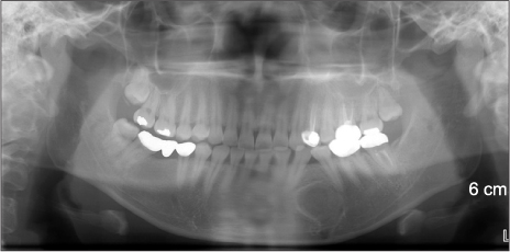
Fig. 3 Post-operative panoramic radiography.

Fig. 4 Deep-stained calcifying materials and eosinophilic amyloids. The epithelial cells shaped like strands surround the calcifying materials. The amyloids show red color in Congo red staining and bright apple green color- which means bi-refringence-in polarizing microscopic view. A. Optical microscopic view (H&E staining, ×40). B. Optical microscopic view (H&E staining, ×200). C. Optical microscopic view (Congo red staining, ×200). D. Polarizing microscopic view (Congo red staining, ×200).
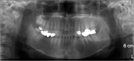
Fig. 5 Post-operative 10-month panoramic radiography.
Reference
-
1. Cheng YS, Wright JM, Walstad WR, Finn MD. Calcifying epithelial odontogenic tumor showing microscopic features of potential malignant behavior. Oral Surg Oral Med Oral Pathol Oral Radiol Endod. 2002. 93:287–295.
Article2. Rapidis AD, Stavrianos SD, Andressakis D, Lagogiannis G, Bertin PM. Calcifying epithelial odontogenic tumor (CEOT) of the mandible: clinical therapeutic conference. J Oral Maxillofac Surg. 2005. 63:1337–1347.
Article3. Philipsen HP, Reichart PA. Calcifying epithelial odontogenic tumour: biological profile based on 181 cases from the literature. Oral Oncol. 2000. 36:17–26.
Article4. Neville BW, Damm DD, Allen CM, Bouquot JE. Oral and maxillofacial pathology. 2002. 3rd ed. Philadelphia: W. B. Saunders.5. Deboni MC, Naclério-Homem Mda G, Pinto DS Junior, Traina AA, Cavalcanti MG. Clinical, radiological and histological features of calcifying epithelial odontogenic tumor: case report. Braz Dent J. 2006. 17:171–174.
Article6. Gon F. The calcifying epithelial odontogenic tumor: report of a case and a study of its histogenesis. Br J Cancer. 1965. 19:39–50.7. Takeda Y, Suzuki A, Sekiyama S. Peripheral calcifying epithelial odontogenic tumor. Oral Surg Oral Med Oral Pathol. 1983. 56:71–75.
Article8. Gopalakrishnan R, Simonton S, Rohrer MD, Koutlas IG. Cystic variant of calcifying epithelial odontogenic tumor. Oral Surg Oral Med Oral Pathol Oral Radiol Endod. 2006. 102:773–777.
Article9. Franklin CD, Pindborg JJ. The calcifying epithelial odontogenic tumor. A review and analysis of 113 cases. Oral Surg Oral Med Oral Pathol. 1976. 42:753–765.10. Anavi Y, Kaplan I, Citir M, Calderon S. Clear-cell variant of calcifying epithelial odontogenic tumor: clinical and radiographic characteristics. Oral Surg Oral Med Oral Pathol Oral Radiol Endod. 2003. 95:332–339.
Article11. Kumamoto H, Sato I, Tateno H, Yokoyama J, Takahashi T, Ooya K. Clear cell variant of calcifying epithelial odontogenic tumor (CEOT) in the maxilla: report of a case with immunohistochemical and ultrastructural investigations. J Oral Pathol Med. 1999. 28:187–191.12. Schmidt-Westhausen A, Philipsen HP, Reichart PA. Clear cell calcifying epithelial odontogenic tumor. A case report. Int J Oral Maxillofac Surg. 1992. 21:47–49.13. Sapp JP, Eversole LR, Wysocki GP. Contemporary oral and maxillofacial pathology. 2004. 2nd ed. New York: Elsevier.14. Damm DD, White DK, Drummond JF, Poindexter JB, Henry BB. Combined epithelial odontogenic tumor: adenomatoid odontogenic tumor and calcifying epithelial odontogenic tumor. Oral Surg Oral Med Oral Pathol. 1983. 55:487–496.
Article
- Full Text Links
-
- Actions
-
Cited
- CITED
-
- Close
- Share
-
- Similar articles
-
- The Calcifying Epithelial Odonogenic Tumor: Report of a Case
- Odontogenic ghost cell carcinoma arising from odontogenic epithelial tumor in maxilla: A case report
- Dentinogenic Ghost Cell Tumor: A Case Report and Review of Literature
- Current Concepts and Occurrence of Epithelial Odontogenic Tumors: II. Calcifying Epithelial Odontogenic Tumor Versus Ghost Cell Odontogenic Tumors Derived from Calcifying Odontogenic Cyst
- Calcifying epithelial odontogenic tumor (CEOT) in palate: report of a case
