Rosai-Dorfman Disease Occurred in Epidural Space of the Thoracic Spinal Canal
- Affiliations
-
- 1Department of Orthopaedic Surgery, Soonchunhyang University College of Medicine, Spine center Seoul, Korea. yil400@hosp.sch.ac.kr
- 2Department of Anatomical Pathology, Soonchunhyang University College of Medicine, Spine center Seoul, Korea.
- 3Department of Orthopaedic Surgery,Soonchunhyang University College of Medicine, Gumi hospital, Gumi, Korea.
- KMID: 2040783
- DOI: http://doi.org/10.4184/jkss.2006.13.1.64
Abstract
- Rosai-Dorfman disease is a rare, non-neoplastic lymphoproliferative disorder that is characterized by its specific histological features. However, it is uncommon for it to involve the thoracic spinal cord as a site of extranodal disease. A 36 year-old man developed progressive paraparesis 2 weeks prior to admission. On an MRI study, the spinal cord was compressed at the T4 and T5 levels posteriorly by an epidural mass. A decompressive laminectomy and removal of the mass were performed and Rosai-Dorfman disease was confirmed histologically. After the operation, additional high-dose radiotherapy was performed. The motor weakness and hypesthesia in the lower extremities resolved completely and there was no evidence of recurrence at the final follow-up examination.
MeSH Terms
Figure
-

Fig. 1. (A)Sagittal T2-weighted magnetic resonance images of thoracic spine showing extradural mass lesion extending from T2, T3, T4 area. (B)Axial T2-weighted magnetic resonance images at the level of 4th thoracic spine showing extradural mass(arrows) and compression of the cord from central to left side.
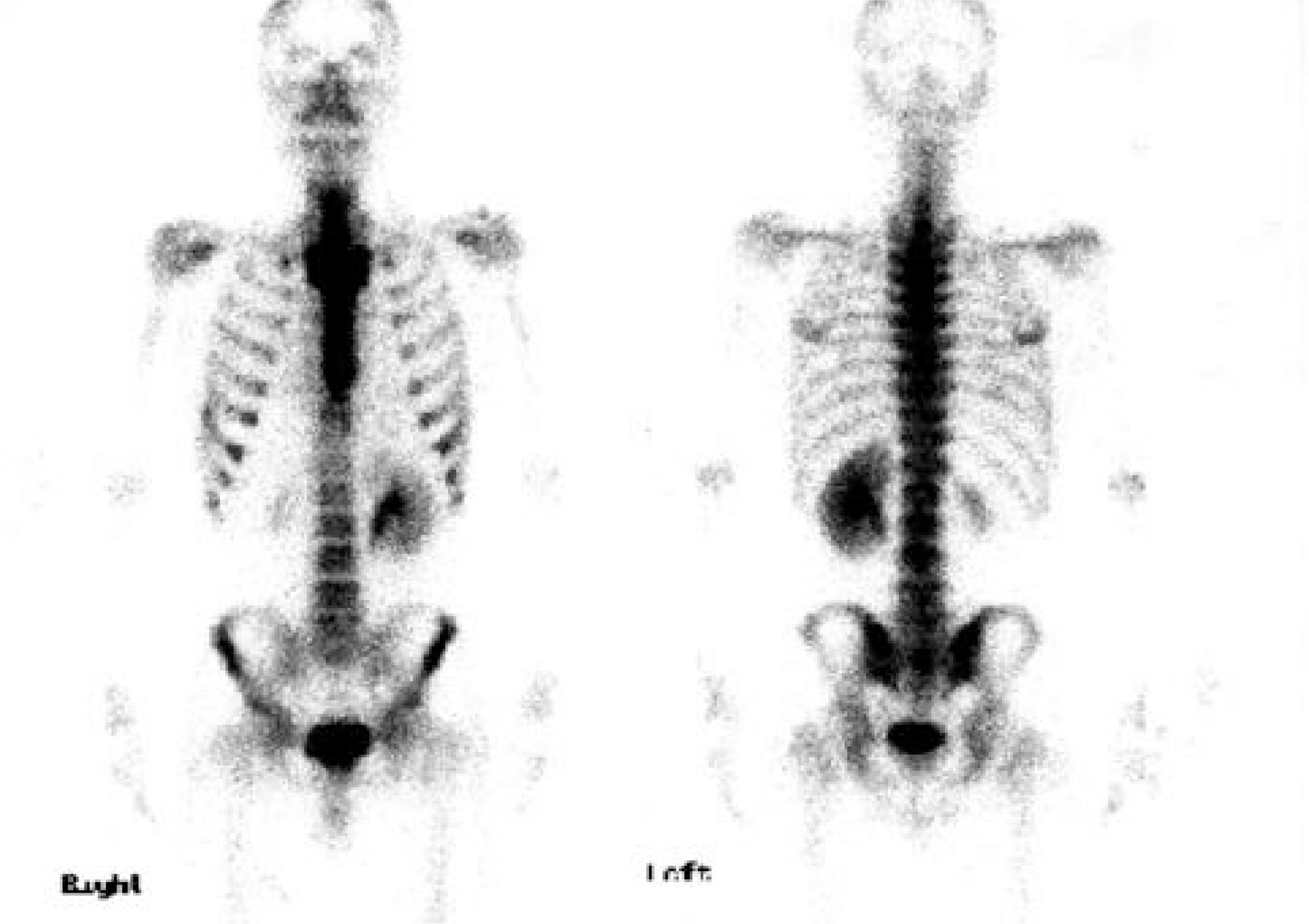
Fig. 2. Preoperative bone scan showing tear drop shaped increased uptake of upper thoracic spine from T1 to T4.
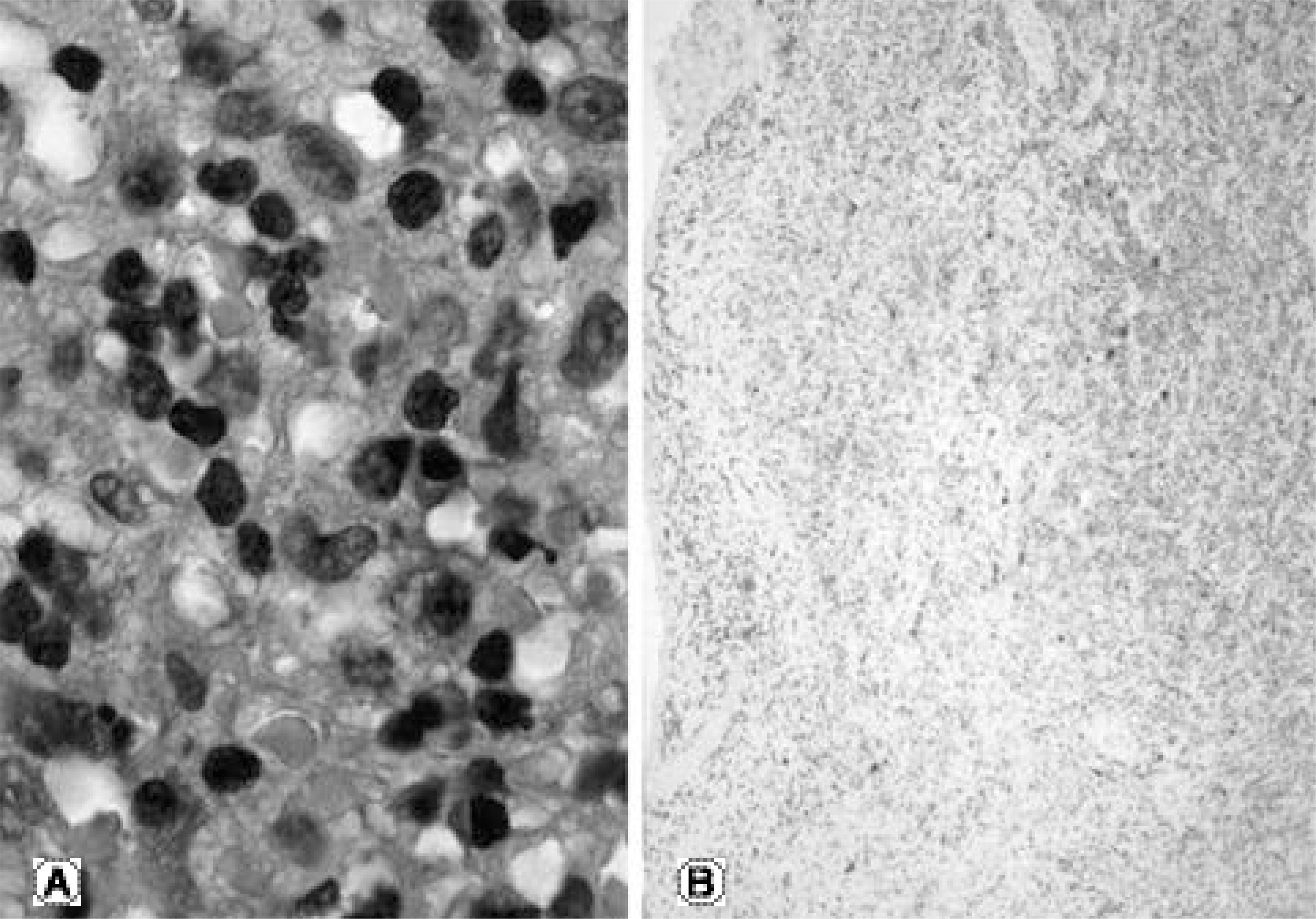
Fig. 3. Histologic findings. (A)A large numbers of histiocytes are interrupted by lymphocytes and plasma cells (B)The histiocytes are positive for S-100 protein.
Cited by 1 articles
-
Rosai-Dorfman Disease as a Solitary Lesion of the Tibia
Chang-Bae Kong, Jung-Wook Lee, Sang-Hyun Cho, Won-Seok Song, Wan-Hyeong Cho, Jae-Soo Koh, Dae-Geun Jeon, Soo-Yong Lee
J Korean Bone Joint Tumor Soc. 2014;20(1):32-35. doi: 10.5292/jkbjts.2014.20.1.32.
Reference
-
1). Sartoris DJ, Resnick D. Osseous involvement in sinus histiocytosis with massive lymphadenopathy (Rosai-Dorfman disease). Eur J Pediatr. 1986; 145:238–40.
Article2). Park JK, Cho MY, Park KH Pyon JS. Isolated intracranial Rosai-Dorfman disease - a case report. J Kor Pathol. 2004; 38:430–433.3). George J, Stacy G, Peabody T, Montag A. Rosai-Dorfman disease manifesting as a solitary lesion of the radius in a 41-year-old woman. Skeletal Radiol. 2003; 32:236–239.
Article4). Raveenthiran V, Dhanalakshmi M, Hayavadana Rao PV, Viswanathan P. Rosai-Dorfman disease: report of a 3-year-old girl with critical review of treatment options. Eur J Pediatr Surg. 2003; 13:350–354.
Article5). Hargett C, Bassett T. Atypical presentation of sinus histiocytosis with massive lymphadenopathy as an epidural spinal cord tumor: a case presentation and literature review. J Spinal Disord Tech. 2005; 18:193–196.6). Geara AR, Ayoubi MA, Achram MC, Chamseddine NM. Rosai-Dorfman disease mimicking neurofibromato -sis: case presentation and review of the literature. Clin Radiol. 2004; 59:625–630.7). Osenbach RK. Isolated extranodal sinus histiocytosis presentimg as an intramedullary spinal cord tumor with paraplegia. J Neurosurg. 1996; 85:692–696.
- Full Text Links
-
- Actions
-
Cited
- CITED
-
- Close
- Share
-
- Similar articles
-
- A Case of Cutaneous Rosai-Dorfman Disease Enlarged in Size after Punch Biopsy
- Rosai-Dorfman Disease in the Neck and Subglottis
- A Case of Rosai-Dorfman Disease Limited to the Lip
- Rapidly Growing Cutaneous Rosai-Dorfman Disease Successfully Treated with Surgical Excision
- Rosai-Dorfman Disease in Thoracic Spine: A Rare Case of Compression Fracture
