J Korean Soc Spine Surg.
2006 Mar;13(1):54-58. 10.4184/jkss.2006.13.1.54.
Gorham's Disease Misdiagnosed as a Simple Compression Fracture in the Thoracic Spine: A Case Report
- Affiliations
-
- 1Department of Orthopaedic Surgery, Soonchunhyang University College of Medicine, Spine center, Seoul Hospital, Seoul, Korea. schsbj@hosp.sch.ac.kr
- 2Department of Orthopaedic Surgery, Soonchunhyang University College of Medicine, Gumi Hospital, Gumi, Korea.
- KMID: 2040781
- DOI: http://doi.org/10.4184/jkss.2006.13.1.54
Abstract
- Gorham's disease is a rare condition of unknown etiology that is characterized by progressive osteolysis. A 48 year-old woman had a burst fracture at T10, which was treated by pedicle screw instrumentation at another hospital. She was transferred due to progressive paraparesis, which was not observed initially. An MRI demonstrated severe cord compression at the T10 level. Under the assumption that the patient had a highly vascular metastatic tumor, an anterior decompression with instrumentation was performed. However, neurologic symptoms and bone destruction worsened after six weeks postoperatively. A repeat decompression was performed through the posterior route and long-level pedicle screw instrumentation was applied. After the second operation, Gorham's disease was confirmed histologically. Care must be taken not to overlook a pathologic fracture caused by a spinal tumor as a simple fracture, especially an osteoporotic one.
Keyword
MeSH Terms
Figure
-

Fig. 1. Initail X-ray showing decreased T10 vertebral body height.
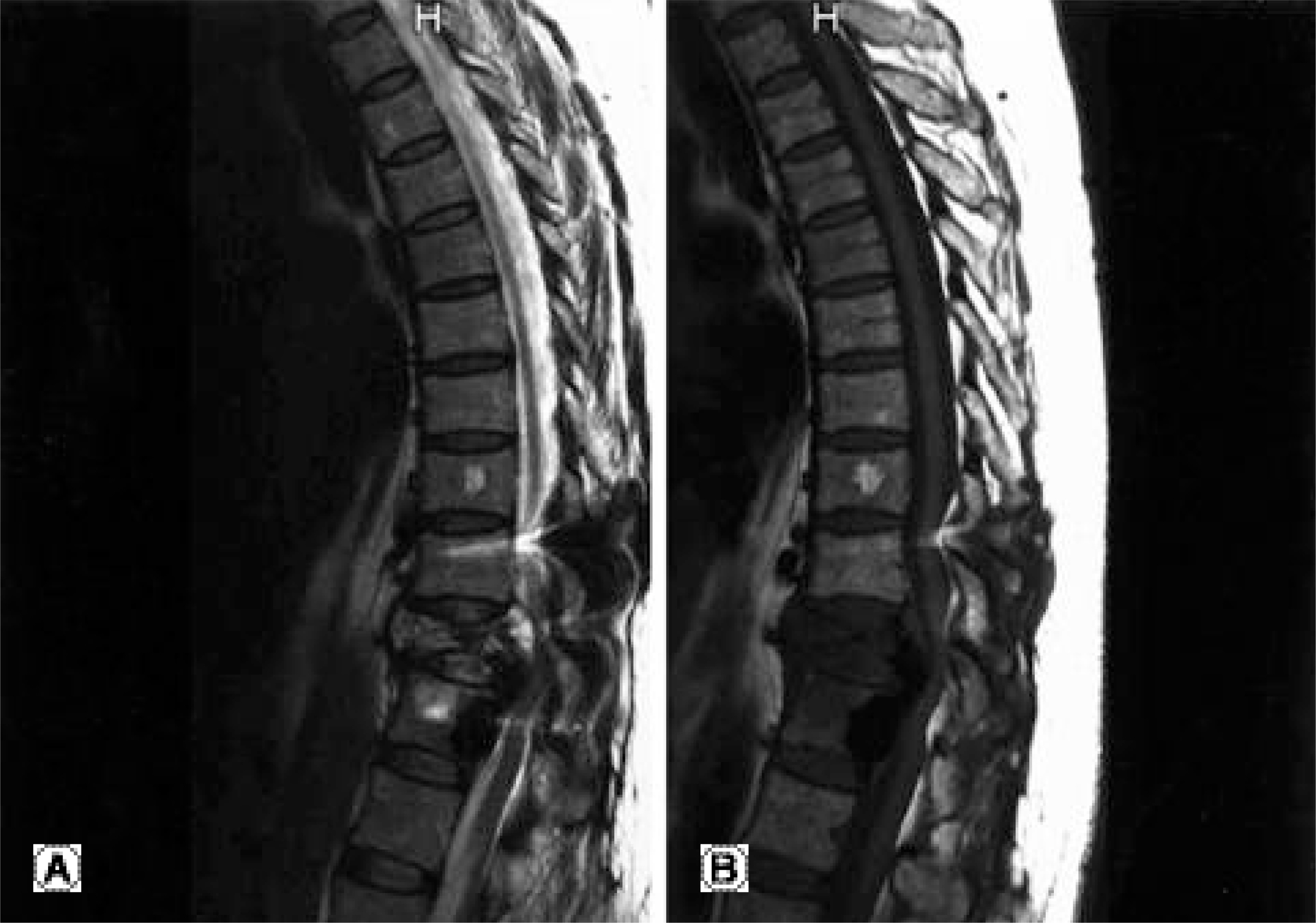
Fig. 2. Preoperative MRI. (A) Sagittal T2-weighted image showing heterogeneous high signal intensities in T10 (B) Sagittal T1-weighted image showing low signal intensities in T10 vertebral body with spinal cord compression.
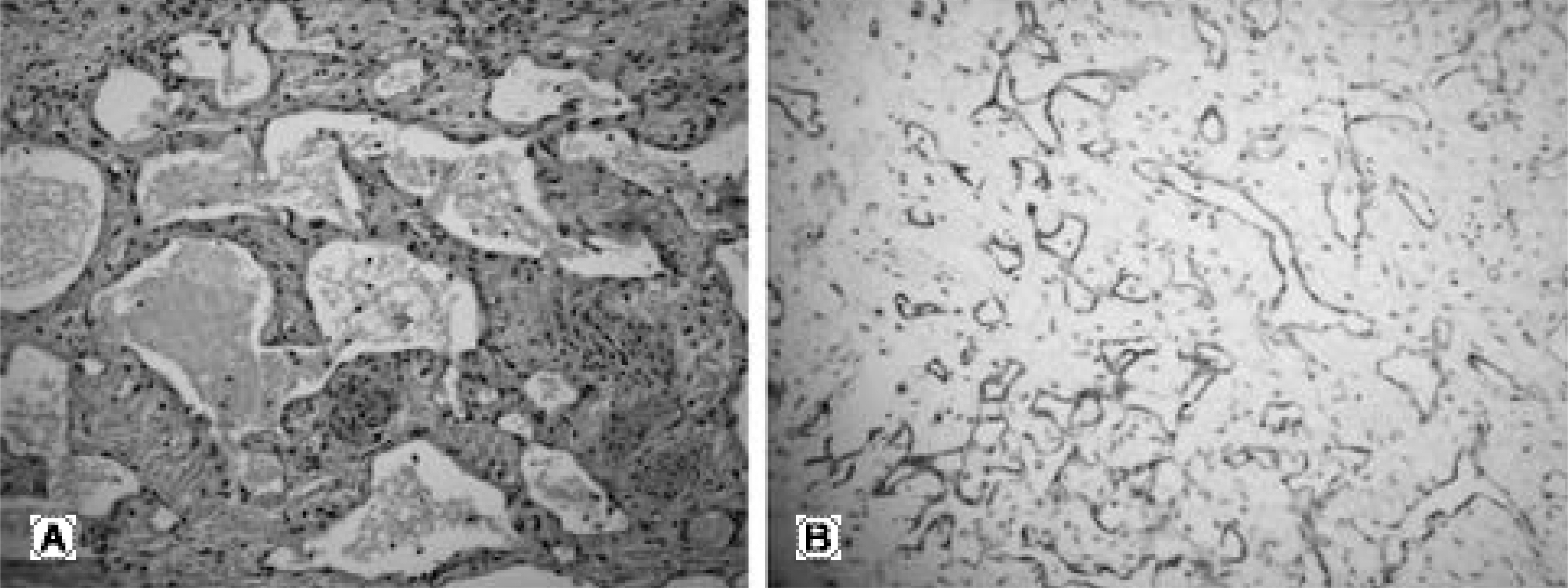
Fig. 3. Histological findings (A) High power view of the tumor. The vascular channels are lined by flat normal appearing endothelial cells without atypia. (H-E× 100). (B) Immunohistochemical finding of the tumor. The lining cells of vascular channels are positive for CD34. (ABC× 100).

Fig. 4. At 1 year postoperatively, radiograph showing no recurrence of Gorham's disease.
Reference
-
1). Gorham LW, Wright AW, Shultz HH, Maxon FC Jr. Disappearing bones. A rare form of massive osteolysis. Am J Med. 1954; 17:674–682.
Article2). Heffez L, Doku HC, Carter BL, Feeney JE. Perspective on massive osteolysis. Report of a case and review of the literature. Oral Surg Oral Med Oral Pathol. 1983; 55:331–343.3). Heyden G, Kindblom LG, Nielsen M. Di sapp earing bone disease. J bone Joint Surg Am. 1977; 59:57–61.4). Jeong BY, Lee CK, Chang BS and Lee DH. Gorham's disease of the spine. J Kor Orthop Assoc. 2002; 37:443–6.5). Johnson PM, McClure JG. Observations on massive osteolysis. A review of the literature and report of a case. Radiology. 1958; 71:28–41.6). Drewry GR, Sutterlin CE, Martinez CR, Brantley SG. Gorham disease of the spine. Spine. 1994; 19:2213–2222.
Article7). Joseph J, Bartal E. Disappearing bone disease. A case report and review of the literature. J Ped Orthop. 1987; 7:584–588.8). Pazzalia UE, Andrini L, Bonato M, Leutner M. Pathology of disappearing bone disease. A case report with immunohistochemical study. Int Orthop. 1997; 21:303–307.9). Choma ND, Biscotti CV, Bauer TW, Metha AC, Licata AA. Gorham's syndrome. A case report and review of the literature. Am J Med. 1987; 83:1151–1156.
Article10). Campbell J, Almond HGA, Johnson R. Massive osteolysis of the humerus with spontaneous recovery. Report of a case. J Bone Joint Surg. Br. 1975; 57:238–240.
- Full Text Links
-
- Actions
-
Cited
- CITED
-
- Close
- Share
-
- Similar articles
-
- Gorham's Disease of Spine
- Comparison of CRP in Spinal Compression Fractures: Simple vs Malignant metastatic disease
- Gorham-Stout Disease in a Middle-Aged Patient Treated by Posterior Lateral Fusion: A Case Report and Literature Review
- Anesthetic experience in a patient with Gorham's disease : A case report
- Gorham's Syndrom: A Case Report
