J Periodontal Implant Sci.
2014 Oct;44(5):222-226. 10.5051/jpis.2014.44.5.222.
Evaluation of alveolar crest bone loss via premolar bitewing radiographs: presentation of a new method
- Affiliations
-
- 1Department of Oral and Maxillofacial Radiology, School of Dentistry, Shahid Beheshti University of Medical Sciences, Tehran, Iran.
- 2Department of Periodontics, School of Dentistry, Shahid Beheshti University of Medical Sciences, Tehran, Iran.
- 3Endodontics Department, Dental Branch, Islamic Azad University, Tehran, Iran.
- 4Department of Oral and Maxillofacial Surgery, School of Dentistry, Shahid Beheshti University of Medical Sciences, Tehran, Iran. esmaeelnejad@gmail.com
- 5Department of Oral and Maxillofacial Pathology, School of Dentistry, Shahid Beheshti University of Medical Sciences, Tehran, Iran.
- KMID: 2027819
- DOI: http://doi.org/10.5051/jpis.2014.44.5.222
Abstract
- PURPOSE
This study aimed to assess the interdental bone level in premolar bitewing radiographs while retracting the cheeks.
METHODS
Seventy-two horizontal bone defects were created on dried mandibles and maxillae. The distance from the bone level to the cement-enamel junction of premolars was detected by a modified digital caliper (considered the gold standard). The reliability of all radiographs was assessed by intraclass correlation coefficient (ICC), and the validity was compared to the gold standard using the analysis of variance test. P-values less than 0.05 were considered statistically significant.
RESULTS
This study showed that the reliability of radiographs without a cheek simulator and with 0.16 second exposure time was significantly higher than that of the two other groups (ICC=0.96 compared to 0.93 and 0.88, respectively). The results from the radiographs without a cheek simulator and with 0.16 second exposure time were more similar to the gold standard measures than those of the two other groups, although the difference was not statistically significant.
CONCLUSIONS
Retracting the buccal soft tissue plays an important role in increasing the accuracy of radiographs in detecting the interdental alveolar bone level and produces more accurate results than increasing the exposure time, although it does not have a significant role in reliability of results.
Keyword
MeSH Terms
Figure
-
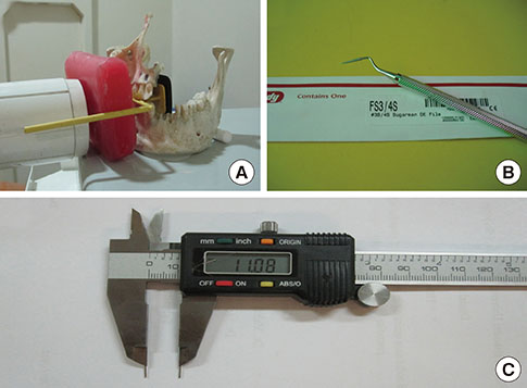
Figure 1 (A) Dried mandible used in the present study. (B) Bone file used to create alveolar bone defects. (C) Digital caliper used to measure the depth of alveolar defects. Two 0.6-mm titanium wires were soldered to the tip of the device to measure the interproximal site accurately.
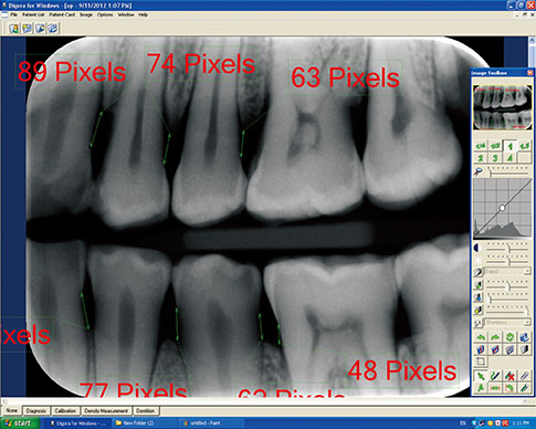
Figure 2 DIGORA software (Soredex, Tuusula, Finland) used for evaluating the radiologic measurements. The distance between the cement-enamel junction and alveolar crest was determined based on the pixel number. Then the pixel value was converted to millimeters.
Reference
-
1. Novak MJ. Classification of diseases and conditions affecting the periodontium. In : Newman MG, Takaei HH, Klokkevold PR, Carranza FA, editors. Carranza's clinical periodontology. 11th ed. St. Louis: Elsevier Saunders Inc.;2012. p. 34–54.2. Armitage GC. Development of a classification system for periodontal diseases and conditions. Ann Periodontol. 1999; 4:1–6.
Article3. Flemmig TF. Periodontitis. Ann Periodontol. 1999; 4:32–38.
Article4. Benn DK. A review of the reliability of radiographic measurements in estimating alveolar bone changes. J Clin Periodontol. 1990; 17:14–21.
Article5. Pepelassi EA, Diamanti-Kipioti A. Selection of the most accurate method of conventional radiography for the assessment of periodontal osseous destruction. J Clin Periodontol. 1997; 24:557–567.
Article6. Kiliç AR, Efeoglu E, Yilmaz S. A new method for standardization of intraoral radiographs. Periodontal Clin Investig. 1996; 18:20–26.7. Gedik R, Marakoglu I, Demirer S. Assessment of alveolar bone levels from bitewing, periapical and panoramic radiographs in periodontitis patients. West Indian Med J. 2008; 57:410–413.8. Kim TS, Obst C, Zehaczek S, Geenen C. Detection of bone loss with different x-ray techniques in periodontal patients. J Periodontol. 2008; 79:1141–1149.
Article9. Pepelassi EA, Tsiklakis K, Diamanti-Kipioti A. Radiographic detection and assessment of the periodontal endosseous defects. J Clin Periodontol. 2000; 27:224–230.
Article10. Vandenberghe B, Jacobs R, Yang J. Diagnostic validity (or acuity) of 2D CCD versus 3D CBCT-images for assessing periodontal breakdown. Oral Surg Oral Med Oral Pathol Oral Radiol Endod. 2007; 104:395–401.
Article11. Eickholz P, Riess T, Lenhard M, Hassfeld S, Staehle HJ. Digital radiography of interproximal bone loss; validity of different filters. J Clin Periodontol. 1999; 26:294–300.
Article12. Kim TS, Benn DK, Eickholz P. Accuracy of computer-assisted radiographic measurement of interproximal bone loss in vertical bone defects. Oral Surg Oral Med Oral Pathol Oral Radiol Endod. 2002; 94:379–387.
Article13. Tihanyi D, Gera I, Eickholz P. Influence of individual brightness and contrast adjustment on accuracy of radiographic measurements of infrabony defects. Dentomaxillofac Radiol. 2011; 40:177–183.
Article14. Horwitz J, Machtei EE, Reitmeir P, Holle R, Kim TS, Eickholz P. Radiographic parameters as prognostic indicators for healing of class II furcation defects. J Clin Periodontol. 2004; 31:105–111.
Article15. Gomes-Filho IS, Sarmento VA, de Castro MS, da Costa NP, da Cruz SS, Trindade SC, et al. Radiographic features of periodontal bone defects: evaluation of digitized images. Dentomaxillofac Radiol. 2007; 36:256–262.
Article16. Benn DK. Frequent, low-dose, improved-contrast radiographic images with the use of narrow x-ray beams. Oral Surg Oral Med Oral Pathol. 1992; 74:221–229.
Article17. Bohay RN. The sensitivity, specificity, and reliability of radiographic periapical diagnosis of posterior teeth. Oral Surg Oral Med Oral Pathol Oral Radiol Endod. 2000; 89:639–642.
Article18. Torres MG, Santos Ada S, Neves FS, Arriaga ML, Campos PS, Crusoé-Rebello I. Assessment of enamel-dentin caries lesions detection using bitewing PSP digital images. J Appl Oral Sci. 2011; 19:462–468.
Article19. Fleiss JL. The design and analysis of clinical experiments. New York: Wiley;1986.20. Kay SM. Fundamentals of statistical signal processing, Volume III: practical algorithm development. Upper Saddle River, NJ: Prentice Hall;2013.21. Schropp L, Alyass NS, Wenzel A, Stavropoulos A. Validity of wax and acrylic as soft-tissue simulation materials used in in vitro radiographic studies. Dentomaxillofac Radiol. 2012; 41:686–690.
Article22. Feijo CV, Lucena JG, Kurita LM, Pereira SL. Evaluation of cone beam computed tomography in the detection of horizontal periodontal bone defects: an in vivo study. Int J Periodontics Restorative Dent. 2012; 32:e162–e168.23. Zybutz M, Rapoport D, Laurell L, Persson GR. Comparisons of clinical and radiographic measurements of inter-proximal vertical defects before and 1 year after surgical treatments. J Clin Periodontol. 2000; 27:179–186.
Article24. Albandar JM, Abbas DK, Waerhaug M, Gjermo P. Comparison between standardized periapical and bitewing radiographs in assessing alveolar bone loss. Community Dent Oral Epidemiol. 1985; 13:222–225.
Article25. Ordinola-Zapata R, Bramante CM, Duarte MH, Ramos Fernandes LM, Camargo EJ, de Moraes IG, et al. The influence of cone-beam computed tomography and periapical radiographic evaluation on the assessment of periapical bone destruction in dog's teeth. Oral Surg Oral Med Oral Pathol Oral Radiol Endod. 2011; 112:272–279.
Article26. de Faria Vasconcelos K, Evangelista KM, Rodrigues CD, Estrela C, de Sousa TO, Silva MA. Detection of periodontal bone loss using cone beam CT and intraoral radiography. Dentomaxillofac Radiol. 2012; 41:64–69.
Article27. Mohan R, Singh A, Gundappa M. Three-dimensional imaging in periodontal diagnosis - Utilization of cone beam computed tomography. J Indian Soc Periodontol. 2011; 15:11–17.
Article28. Esmaeli F, Shirmohammadi A, Faramarzie M, Abolfazli N, Rasouli H, Fallahi S. Determination of vertical interproximal bone loss topography: correlation between indirect digital radiographic measurement and clinical measurement. Iran J Radiol. 2012; 9:83–87.
Article29. Henriksson CH, Stermer EM, Aass AM, Sandvik L, Moystad A. Comparison of the reproducibility of storage phosphor and film bitewings for assessment of alveolar bone loss. Acta Odontol Scand. 2008; 66:380–384.
Article30. Wolf B, von Bethlenfalvy E, Hassfeld S, Staehle HJ, Eickholz P. Reliability of assessing interproximal bone loss by digital radiography: intrabony defects. J Clin Periodontol. 2001; 28:869–878.
Article
- Full Text Links
-
- Actions
-
Cited
- CITED
-
- Close
- Share
-
- Similar articles
-
- A study about alveolar crest bone height before and after orthodontic treatment by using bitewing film
- An evaluation of angles between the alveolar crest bone and the implant effect on the implant crestal area induced stresses using a finite element method
- Relationship between Interdental Papilla Existence & Distance from Interdental Alveolar Crest to Contact Point in the Posterior Dentition of Korean adults
- Prediction of the alveolar bone level after the extraction of maxillary anterior teeth with severe periodontitis
- A study on the change of alveolar crest height following orthodontic treatment
