J Periodontal Implant Sci.
2015 Dec;45(6):216-222. 10.5051/jpis.2015.45.6.216.
Prediction of the alveolar bone level after the extraction of maxillary anterior teeth with severe periodontitis
- Affiliations
-
- 1Department of Periodontology, Pusan National University School of Dentistry, Yangsan, Korea. borum2@hanmail.net
- 2Department of Periodontology, Pusan National University Dental Hospital, Dental Research Institute, Yangsan, Korea.
- KMID: 2354939
- DOI: http://doi.org/10.5051/jpis.2015.45.6.216
Abstract
- PURPOSE
After extraction, the alveolar bone tends to undergo atrophy in three-dimensions. The amount of alveolar bone loss in the horizontal dimension has been reported to be greater than the amount of bone loss in the vertical dimension, and is most pronounced in the buccal aspect. The aim of this study was to monitor the predictive alveolar bone level following the extraction of anterior teeth seriously involved with advanced chronic periodontitis.
METHODS
This study included 25 patients with advanced chronic periodontitis, whose maxillary anterior teeth had been extracted due to extensive attachment loss more than one year before the study. Periapical radiographs were analyzed to assess the vertical level of alveolar bone surrounding the edentulous area. An imaginary line connecting the mesial and the distal ends of the alveolar crest facing the adjacent tooth was arbitrarily created. Several representative coordinates were established in the horizontal direction, and the vertical distance from the imaginary line to the alveolar crest was measured at each coordinate for each patient using image analysis software. Regression functions predicting the vertical level of the alveolar bone in the maxillary anterior edentulous area were identified for each patient.
RESULTS
The regression functions demonstrated a tendency to converge to parabolic shapes. The predicted maximum distance between the imaginary line and the alveolar bone calculated using the regression function was 1.43+/-0.65 mm. No significant differences were found between the expected and actual maximum distances. Likewise, the predicted and actual maximum horizontal distances did not show any significant differences. The distance from the alveolar bone crest to the imaginary lines was not influenced by the mesio-distal spans of the edentulous area.
CONCLUSIONS
After extraction, the vertical level of the alveolar ridge increased to become closer to the reference line connecting the mesial and distal alveolar crests.
Keyword
MeSH Terms
Figure
-
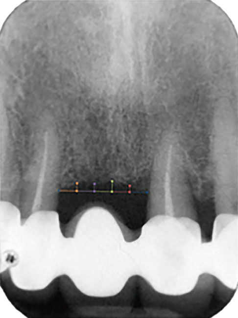
Figure 1 An imaginary line connecting the mesial and the distal ends of the alveolar crest facing each adjacent tooth was created. The distances between the imaginary line and several representative points were measured for each patient using an image analysis program.
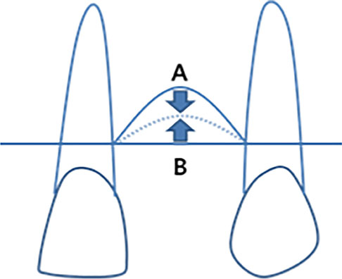
Figure 2 Convergence line indicating the vertical level of the alveolar crest after extraction. A: Change in the vertical level of the alveolar crest after extraction of teeth with severe bone loss. B: Change in the vertical level of the alveolar crest after extraction of teeth with a normal bone level.
Reference
-
1. Tallgren A. The continuing reduction of the residual alveolar ridges in complete denture wearers: a mixed-longitudinal study covering 25 years. 1972. J Prosthet Dent. 2003; 89:427–462.2. Pinho MN, Roriz VL, Novaes AB Jr, Taba M Jr, Grisi MF, de Souza SL, et al. Titanium membranes in prevention of alveolar collapse after tooth extraction. Implant Dent. 2006; 15:53–61.
Article3. Kerr EN, Mealey BL, Noujeim ME, Lasho DJ, Nummikoski PV, Mellonig JT. The effect of ultrasound on bone dimensional changes following extraction: a pilot study. J Periodontol. 2008; 79:283–373.
Article4. Darby I, Chen ST, Buser D. Ridge preservation techniques for implant therapy. Int J Oral Maxillofac Implants. 2009; 24:Suppl. 260–331.5. Lekovic V, Kenney EB, Weinlaender M, Han T, Klokkevold P, Nedic M, et al. A bone regenerative approach to alveolar ridge maintenance following tooth extraction. Report of 10 cases. J Periodontol. 1997; 68:563–633.
Article6. Lekovic V, Camargo PM, Klokkevold PR, Weinlaender M, Kenney EB, Dimitrijevic B, et al. Preservation of alveolar bone in extraction sockets using bioabsorbable membranes. J Periodontol. 1998; 69:1044–1053.
Article7. Iasella JM, Greenwell H, Miller RL, Hill M, Drisko C, Bohra AA, et al. Ridge preservation with freeze-dried bone allograft and a collagen membrane compared to extraction alone for implant site development: a clinical and histologic study in humans. J Periodontol. 2003; 74:990–999.
Article8. Barone A, Aldini NN, Fini M, Giardino R, Calvo Guirado JL, Covani U. Xenograft versus extraction alone for ridge preservation after tooth removal: a clinical and histomorphometric study. J Periodontol. 2008; 79:1370–1377.
Article9. Camargo PM, Lekovic V, Weinlaender M, Klokkevold PR, Kenney EB, Dimitrijevic B, et al. Influence of bioactive glass on changes in alveolar process dimensions after exodontia. Oral Surg Oral Med Oral Pathol Oral Radiol Endod. 2000; 90:581–587.
Article10. Fiorellini JP, Howell TH, Cochran D, Malmquist J, Lilly LC, Spagnoli D, et al. Randomized study evaluating recombinant human bone morphogenetic protein-2 for extraction socket augmentation. J Periodontol. 2005; 76:605–618.
Article11. Schropp L, Wenzel A, Kostopoulos L, Karring T. Bone healing and soft tissue contour changes following single-tooth extraction: a clinical and radiographic 12-month prospective study. Int J Periodontics Restorative Dent. 2003; 23:313–336.12. Pietrokovski J, Massler M. Alveolar ridge resorption following tooth extraction. J Prosthet Dent. 1967; 17:21–28.
Article13. Jahangiri L, Devlin H, Ting K, Nishimura I. Current perspectives in residual ridge remodeling and its clinical implications: a review. J Prosthet Dent. 1998; 80:224–261.
Article14. Araújo MG, Lindhe J. Dimensional ridge alterations following tooth extraction. An experimental study in the dog. J Clin Periodontol. 2005; 32:212–220.
Article15. Bartold PM, Cantley MD, Haynes DR. Mechanisms and control of pathologic bone loss in periodontitis. Periodontol 2000. 2010; 53:55–69.
Article16. Serino G, Biancu S, Iezzi G, Piattelli A. Ridge preservation following tooth extraction using a polylactide and polyglycolide sponge as space filler: a clinical and histological study in humans. Clin Oral Implants Res. 2003; 14:651–659.
Article17. Brägger U, Schild U, Lang NP. Effect of chlorhexidine (0.12%) rinses on periodontal tissue healing after tooth extraction. (II). Radiographic parameters. J Clin Periodontol. 1994; 21:422–452.
Article
- Full Text Links
-
- Actions
-
Cited
- CITED
-
- Close
- Share
-
- Similar articles
-
- Angulation between Long Axis of Anterior Teeth and Alveolar Process, and Thickness of Alveolar Bone
- Single-tooth implant restoration with alveolar bone augmentation in the maxillary anterior tooth region: a case report
- Minimally traumatic extraction of Dentistry fractured bilateral maxillary canine teeth using a piezoelectric surgical unit in an African lion (Panthera leo)
- Effect of slow forced eruption on the vertical levels of the interproximal bone and papilla and the width of the alveolar ridge
- Healing pattern of the mucous membrane after tooth extraction in the maxillary sinus
