Surgical Treatment of Giant Serpentine Aneurysm of A2-A3 Segment Distal Anterior Cerebral Artery : Technical Case Report
- Affiliations
-
- 1Department of Neurosurgery, Chonnam National University Hospital and Medical School, Gwangju, Korea. nsjsp@jnu.ac.kr
- KMID: 2018262
- DOI: http://doi.org/10.3340/jkns.2012.52.5.501
Abstract
OBJECTIVE
To report our surgical experience using in situ end-to-side bypass for giant serpentine distal anterior cerebral artery aneurysm, unsuitable for microsurgical clipping.
METHODS
A 49-year-old woman presented with headache and intermittent loss of consciousness. The brain computed tomography scan revealed a partially calcified mass in the interhemispheric fissure. On cerebral angiography, that was giant (30x18 mm sized), serpentine aneurysm originating from the A2 to A3 segment of the distal anterior cerebral artery (DACA). The aneurysm was trapped with clips, and the right A3 segment to left A3 segment of DACA, end-to-side in situ bypass was performed. Surgical result was favorable, with no newly developed ischemic event in the acute recovery period. Postoperative angiography showed total occlusion of the aneurysm and good patency, with preserved distal flow.
CONCLUSION
Giant fusiform aneurysms of the DACA are extremely rare and can be particularly challenging to treat. End-to-side A3 : A3 bypass with aneurysm trapping could be a treatment modality for these locations.
Keyword
MeSH Terms
Figure
-

Fig. 1 Non-contrast computed tomography demonstrating a 3×1.8 cm sized, rim calcified, and bilobulated mass lesion in the above corpus callosum. B : Brain T2-weighted magnetic resonance image revealed partially thrombosed, signal voided aneurysm. C : Cerebral angiography showing a 4.5×1.8 cm sized, irregular serpentine aneurysm. D : 3-D angiography image of the aneurysm involving the entire A2 segment of the distal anterior cerebral artery. E : Entire scheme of distal anterior cerebral artery serpentine giant fusiform aneurysm.

Fig. 2 Intraoperative findings. A : 4×4 cm sized bone flap is made and dural flap is reflected after cutting the superior sagittal sinus. B : Atherosclerotic distal end of aneurysm is seen at the top. C : Both olfactory nerves are intact and dissected well. D : End to side bypass. E : Atherosclerotic, calcified aneurysm with small opening for blood aspiration.
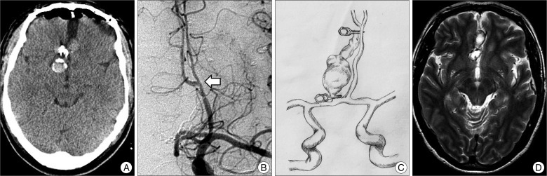
Fig. 3 A : Postoperative computed tomography scan showing a placed clip and no hemorrhagic contusion. B : Follow-up angiography demonstrating good patency of bypass (arrow). C : Follow-up T2-weighted magnetic resonance image showing reduced aneurysm size. D : Follow-up T2-weighted magnetic resonance image showing reduced aneurysm size.
Reference
-
1. Dunn GP, Gerrard JL, Jho DH, Ogilvy CS. Surgical treatment of a large fusiform distal anterior cerebral artery aneurysm with In Situ end-to-side A3-A3 bypass graft and aneurysm trapping : case report and review of the literature. Neurosurgery. 2011; 68:E587–E591. discussion E591. PMID: 21135720.2. Gelfenbeyn M, Natarajan SK, Sekhar LN. Large distal anterior cerebral artery aneurysm treated with resection and interposition graft : case report. Neurosurgery. 2009; 64:E1008–E1009. discussion E1009. PMID: 19404124.3. Ikeda A, Shibuya M, Okada T, Kageyama N. [Microvascular side-to-side anastomosis. Basic problems and clinical applications]. Neurol Med Chir (Tokyo). 1986; 26:379–384. PMID: 2429217.4. Lehecka M, Dashti R, Hernesniemi J, Niemelä M, Koivisto T, Ronkainen A, et al. Microneurosurgical management of aneurysms at the A2 segment of anterior cerebral artery (proximal pericallosal artery) and its frontobasal branches. Surg Neurol. 2008; 70:232–246. discussion 246. PMID: 18486199.
Article5. Lehecka M, Lehto H, Niemelä M, Juvela S, Dashti R, Koivisto T, et al. Distal anterior cerebral artery aneurysms : treatment and outcome analysis of 501 patients. Neurosurgery. 2008; 62:590–601. discussion 590-601. PMID: 18425008.6. Ramanathan D, Hegazy A, Mukherjee SK, Sekhar LN. Intracranial in situ side-to-side microvascular anastomosis : principles, operative technique, and applications. World Neurosurg. 2010; 73:317–325. PMID: 20849786.
Article7. Sanai N, Zador Z, Lawton MT. Bypass surgery for complex brain aneurysms : an assessment of intracranial-intracranial bypass. Neurosurgery. 2009; 65:670–683. discussion 683. PMID: 19834371.
- Full Text Links
-
- Actions
-
Cited
- CITED
-
- Close
- Share
-
- Similar articles
-
- Giant Serpentine Aneurysm of the Anterior Communicating Artery: Case Report
- Giant Serpentine Aneurysm of the Posterior Cerebral Artery: Case Report
- Giant Serpentine Intracranial Aneurysm Treated with Wrapping under the Extracorporeal Circulation and Hypothermia
- Giant Serpentine Aneurysm of the Middle Cerebral Artery
- A2 Anomaly Associated with Anterior Cerebral Artery Aneurysm
