Clear cell odontogenic carcinoma mimicking a cystic lesion: a case of misdiagnosis
- Affiliations
-
- 1Department of Oral and Maxillofacial Surgery, College of Dentistry, Yonsei University, Seoul, Korea. omsnam@yuhs.ac
- 2Department of Oral Pathology, College of Dentistry, Yonsei University, Seoul, Korea.
- 3Oral Cancer Research Institute, College of Dentistry, Yonsei University, Seoul, Korea.
- KMID: 2005446
- DOI: http://doi.org/10.5125/jkaoms.2014.40.4.199
Abstract
- Clear cell odontogenic carcinoma (CCOC) is a rare jaw tumor that was classified as a malignant tumor of odontogenic origin in 2005 by the World Health Organization because of its aggressive and destructive growth capacity and metastasis to the lungs and lymph nodes. We report a case of a 66-year-old female who had swelling, incision and drainage history and a well-defined unicystic radiolucent lesion that was comparable to a cystic lesion. At first, the patient received decompression, and the lesion size decreased. Three months after decompression, cyst enucleation was performed. The pathologic result indicated that the lesion was CCOC. In this report we emphasize that patients with painful cystic lesions in addition to jaw enlargement and loosening teeth should be considered for the possibility of malignancy.
Keyword
MeSH Terms
Figure
-
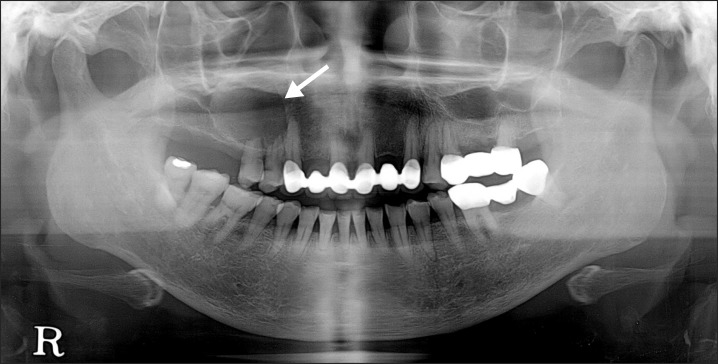
Fig. 1 Panoramic radiograph showing a defined radiolucent lesion in the right maxillary region (arrow).
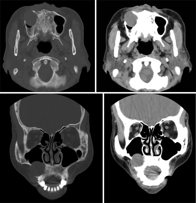
Fig. 2 Computed tomography image at first visit showed well defined and low attenuated cystic lesion on the right maxillary region.
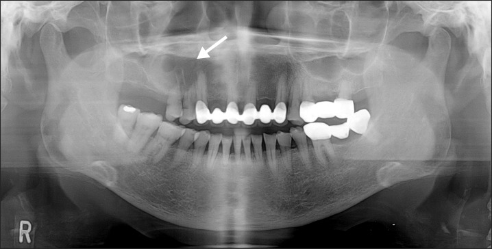
Fig. 3 Two month after decompression, a panoramic radiograph showed that a lesion size decreased, compared with previous radiograph (arrow).
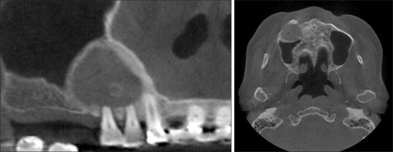
Fig. 4 Three-month follow-up cone-beam computed tomography image after decompression.
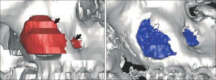
Fig. 5 Three-dimensional reconstruction image, image at first visit is on the left (lesion is indicated with black arrows) and image at 3-months after decompression is on the right (lesion is indicated with white arrows).
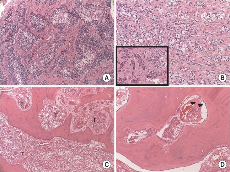
Fig. 6 Histopathologic findings. A. Histopathologic examination of the previous enucleation specimen revealed tumorous proliferation of epithelial nests infiltrating the stroma, indicating malignancy (H&E staining, ×100). B. Cuboidal shaped odontogenic cells were also noted among the tumorous clear cells (H&E staining, B: ×200, inset box: ×200). C. In the resected specimen, the tumor cells (T) infiltrated to the bone marrow of the maxilla (H&E staining, ×100). D. Lymphovascular permeation of tumor cells (arrowheads) (H&E staining, ×200).
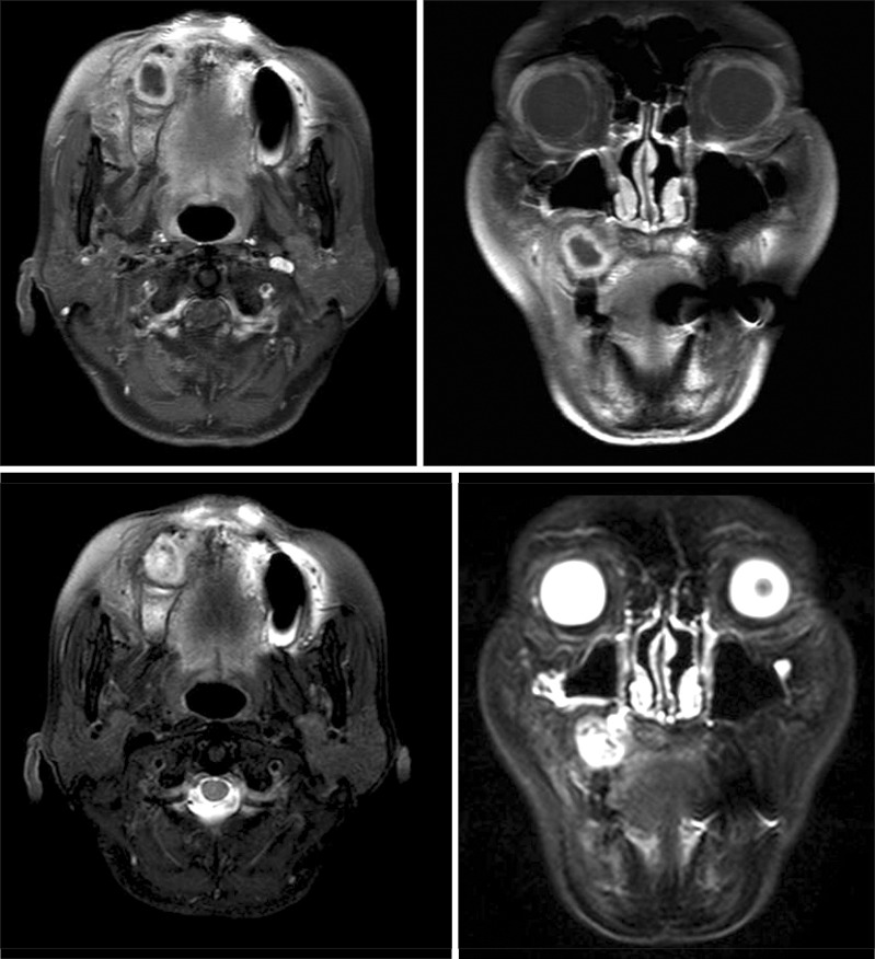
Fig. 7 Magnetic resonance imaging after enucleation of the lesion. The top image is T1-weighted and lower image is T2-weighted. Magnetic resonance images show a well-defined cystic mass.
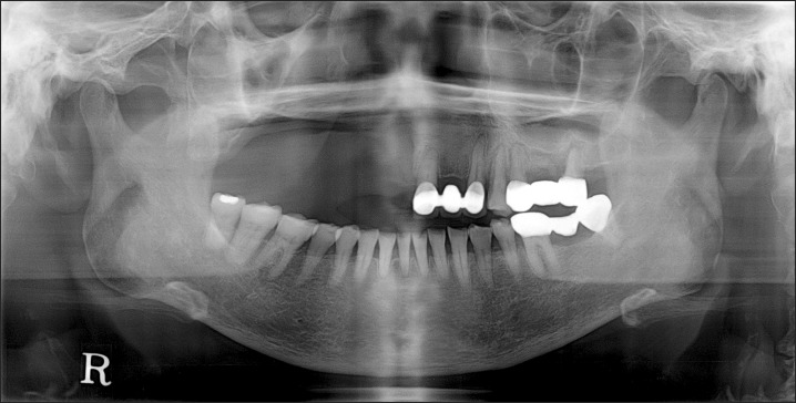
Fig. 8 There was no evidence of local recurrence on eight month follow-up panoramic radiograph after operation.
Cited by 3 articles
-
Misdiagnosis of ameloblastoma in a patient with clear cell odontogenic carcinoma: a case report
Jong-Cheol Park, Seong-Won Kim, Young-Jae Baek, Hyeong-Geun Lee, Mi-Heon Ryu, Dae-Seok Hwang, Uk-Kyu Kim
J Korean Assoc Oral Maxillofac Surg. 2019;45(2):116-120. doi: 10.5125/jkaoms.2019.45.2.116.A radiolucent lesion of the jaw as a presentation form of a mucoepidermoid carcinoma of the oral cavity
David A. Dominguez-Medina, Juan F. Peña-Cardelles, Felix Manzarbeitia-Arambarri
J Korean Assoc Oral Maxillofac Surg. 2021;47(3):229-232. doi: 10.5125/jkaoms.2021.47.3.229.Sequential treatment from mandibulectomy to reconstruction on mandibular oral cancer – Case review II: mandibular anterior and the floor of the mouth lesion of basaloid squamous cell carcinoma and clear cell odontogenic carcinoma
Jae-Young Yang, Dae-Seok Hwang, Uk-Kyu Kim
J Korean Assoc Oral Maxillofac Surg. 2021;47(3):216-223. doi: 10.5125/jkaoms.2021.47.3.216.
Reference
-
1. Hansen LS, Eversole LR, Green TL, Powell NB. Clear cell odontogenic tumor--a new histologic variant with aggressive potential. Head Neck Surg. 1985; 8:115–123. PMID: 4077550.2. Kramer IR, Pindborg JJ, Shear M. Histological typing of odontogenic tumours. Berlin: Springer;1992.3. Barnes L. Pathology and genetics of head and neck tumours. Lyon: IARC Press;2005.4. Kumar M, Fasanmade A, Barrett AW, Mack G, Newman L, Hyde NC. Metastasising clear cell odontogenic carcinoma: a case report and review of the literature. Oral Oncol. 2003; 39:190–194. PMID: 12509974.
Article5. Brinck U, Gunawan B, Schulten HJ, Pinzon W, Fischer U, Füzesi L. Clear-cell odontogenic carcinoma with pulmonary metastases resembling pulmonary meningothelial-like nodules. Virchows Arch. 2001; 438:412–417. PMID: 11355179.
Article6. Piattelli A, Sesenna E, Trisi P. Clear cell odontogenic carcinoma. Report of a case with lymph node and pulmonary metastases. Eur J Cancer B Oral Oncol. 1994; 30B:278–280. PMID: 7950843.
Article7. Fan J, Kubota E, Imamura H, Shimokama T, Tokunaga O, Katsuki T, et al. Clear cell odontogenic carcinoma. A case report with massive invasion of neighboring organs and lymph node metastasis. Oral Surg Oral Med Oral Pathol. 1992; 74:768–775. PMID: 1488233.8. Bang G, Koppang HS, Hansen LS, Gilhuus-Moe O, Aksdal E, Persson PG, et al. Clear cell odontogenic carcinoma: report of three cases with pulmonary and lymph node metastases. J Oral Pathol Med. 1989; 18:113–118. PMID: 2746520.
Article9. Swain N, Dhariwal R, Ray JG. Clear cell odontogenic carcinoma of maxilla: A case report and mini review. J Oral Maxillofac Pathol. 2013; 17:89–94. PMID: 23798837.
Article10. Zhang J, Liu L, Pan J, Tian X, Tan J, Zhou J, et al. Clear cell odontogenic carcinoma: report of 6 cases and review of the literature. Med Oncol. 2011; 28(Suppl 1):S626–S633. PMID: 20827579.
Article11. Werle H, Blake FA, Reichelt U, Schmelzle R, Heiland M. Clear-cell odontogenic carcinoma: a new case and long-term follow-up of an old case, and review of the literature. J Oral Maxillofac Surg. 2009; 67:1342–1348. PMID: 19446231.
Article12. Ebert CS Jr, Dubin MG, Hart CF, Chalian AA, Shockley WW. Clear cell odontogenic carcinoma: a comprehensive analysis of treatment strategies. Head Neck. 2005; 27:536–542. PMID: 15772956.
Article13. Avninder S, Rakheja D, Bhatnagar A. Clear cell odontogenic carcinoma: a diagnostic and therapeutic dilemma. World J Surg Oncol. 2006; 4:91. PMID: 17156493.
Article14. Chaine A, Pitak-Arnnop P, Dhanuthai K, Bertrand JC, Bertolus C. An asymptomatic radiolucent lesion of the maxilla. Clear cell odontogenic carcinoma. Oral Surg Oral Med Oral Pathol Oral Radiol Endod. 2009; 107:452–457. PMID: 19071036.
- Full Text Links
-
- Actions
-
Cited
- CITED
-
- Close
- Share
-
- Similar articles
-
- Ghost Cell Odontogenic Carcinoma Arising from Calcifying Cystic Odontogenic Tumor: A Case Report
- Misdiagnosis of ameloblastoma in a patient with clear cell odontogenic carcinoma: a case report
- Squamous cell carcinoma arising from residual odontogenic cyst: Report of a Case & Review of Literatures
- Multiloculated Cystic Type Renal Epithelioid Angiomyolipoma Mimicking Renal Cell Carcinoma: A Case Report
- Multilocular Cystic Renal Cell Carcinoma: A case report
