Preliminary evaluation of a three-dimensional, customized, and preformed titanium mesh in peri-implant alveolar bone regeneration
- Affiliations
-
- 1Department of Periodontology, Ewha Womans University Mokdong Hospital, Seoul, Korea.
- 2Division of Oral and Maxillofacial Surgery, Department of Dentistry, Hanyang University College of Medicine, Seoul, Korea. fastchang@hanyang.ac.kr
- KMID: 2005443
- DOI: http://doi.org/10.5125/jkaoms.2014.40.4.181
Abstract
OBJECTIVES
The purpose of this preliminary study is to evaluate the effectiveness of a customized, three-dimensional, preformed titanium mesh as a barrier membrane for peri-implant alveolar bone regeneration.
MATERIALS AND METHODS
Ten patients were recruited for this study. At the time of implant placement, all patients had fenestration or a dehiscence defect around the implant fixture. A mixture of particulate intraoral autologous bone and freeze-dried bone allograft was applied to the defect in a 1 : 1 volume ratio and covered by the preformed titanium mesh. A core biopsy specimen was taken from the regenerated bone four months postoperatively. Patients were followed for 12 months after the definitive prosthesis was placed.
RESULTS
Satisfactory bone regeneration with limited fibrous tissue was detected beneath the preformed titanium mesh. Histologic findings revealed that newly formed bones were well-incorporated into the allografts and connective tissue. New growth was composed of approximately 80% vital bone, 5% fibrous marrow tissue, and 15% remaining allograft. All implants were functional without any significant complications.
CONCLUSION
The use of preformed titanium mesh may support bone regeneration by maintaining space for new bone growth through its macro-pores. This preliminary study presents the efficacy of a preformed titanium mesh as a ready-to-use barrier membrane around peri-implant alveolar bone defect. This preformed mesh is also convenient to apply and to remove.
Keyword
MeSH Terms
Figure
-
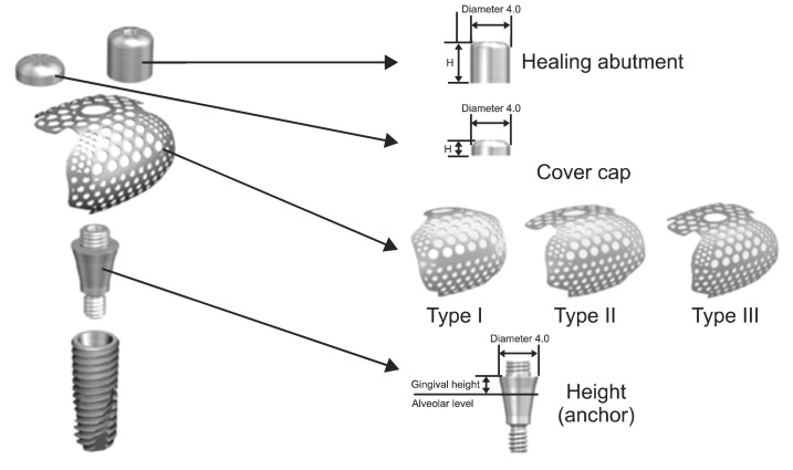
Fig. 1 Components and types of the customized, three-dimensional, and preformed titanium mesh (SMARTbuilder; Osstem). Type I is designed for 1-wall augmentation, type II for 2-wall augmentation, and type III for 3-wall augmentation.
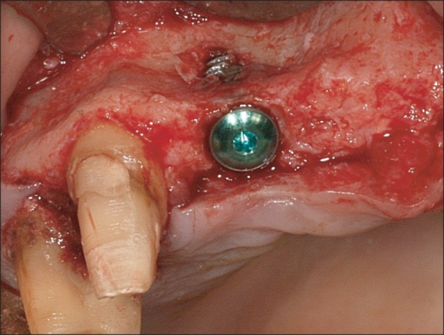
Fig. 2 After implant placement on #12 missing area, labial fenestration defect was found.
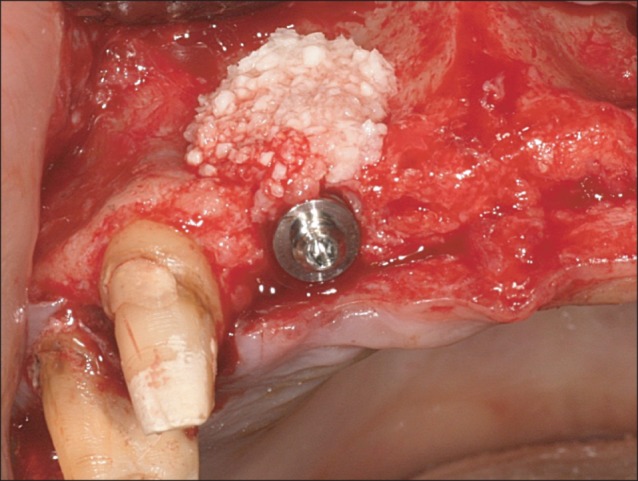
Fig. 3 After connecting the height on the implant fixture, the defect was covered with a mixture of particulated autologous bone harvested from the mandibular ramus and freeze-dried bone allograft.
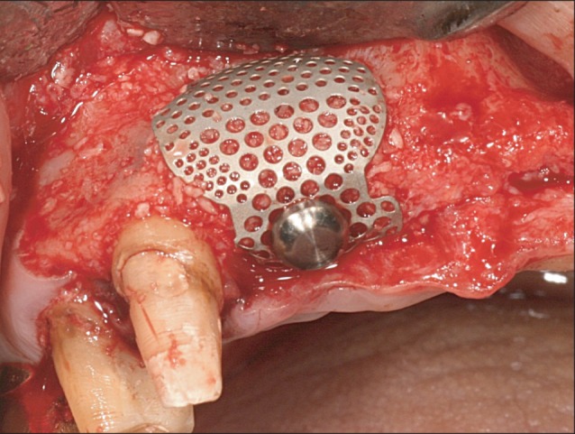
Fig. 4 A customized, 3-dimensional, and preformed titanium mesh (SMARTbuilder type II; Osstem) was connected to the height, placed over the graft material, and stabilized by the cover cap.
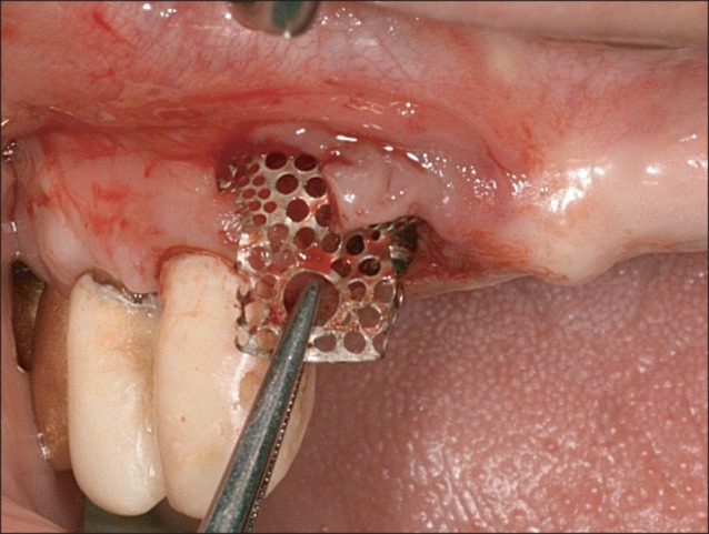
Fig. 5 The preformed titanium mesh was easily removed under the minimum flap elevation at postoperatively 4-month re-entry.
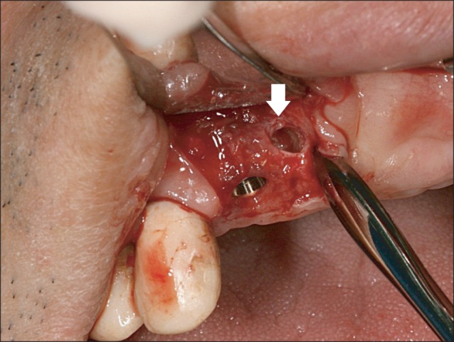
Fig. 6 Bone core biopsy was carried out on the labial regenerated bone by a trephine drill (arrow). Note that there was a superficial thin fibrous tissue layer over the regenerated bone.
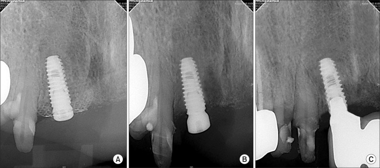
Fig. 7 Comparison of periapical radiographs taken at the application (A) and removal (B) of the preformed titanium mesh and at 1 year after delivery of the final prosthesis (C).
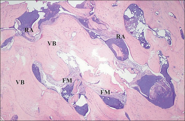
Fig. 8 Histological findings of the core specimen obtained from patient #2. Note that the residual allograft (RA) particles were encapsulated and incorporated with vital bone (VB). Fibrous marrow (FM) tissues were also observed (H&E staining, ×40).
Reference
-
1. Brånemark PI, Zarb GA, Albrektsson T. Tissue-integrated prostheses: osseointegration in clinical dentistry. Chicago: Quintessence;1985. p. 199–209.2. Misch CM. Comparison of intraoral donor sites for onlay grafting prior to implant placement. Int J Oral Maxillofac Implants. 1997; 12:767–776. PMID: 9425757.3. Hämmerle CH, Karring T. Guided bone regeneration at oral implant sites. Periodontol 2000. 1998; 17:151–175. PMID: 10337322.
Article4. Simion M, Baldoni M, Zaffe D. Jawbone enlargement using immediate implant placement associated with a split-crest technique and guided tissue regeneration. Int J Periodontics Restorative Dent. 1992; 12:462–473. PMID: 1298734.5. Oda T, Sawaki Y, Ueda M. Experimental alveolar ridge augmentation by distraction osteogenesis using a simple device that permits secondary implant placement. Int J Oral Maxillofac Implants. 2000; 15:95–102. PMID: 10697943.6. Rakhmatia YD, Ayukawa Y, Furuhashi A, Koyano K. Current barrier membranes: titanium mesh and other membranes for guided bone regeneration in dental applications. J Prosthodont Res. 2013; 57:3–14. PMID: 23347794.
Article7. Boyne PJ. Restoration of osseous defects in maxillofacial casualities. J Am Dent Assoc. 1969; 78:767–776. PMID: 4975262.8. Corinaldesi G, Pieri F, Sapigni L, Marchetti C. Evaluation of survival and success rates of dental implants placed at the time of or after alveolar ridge augmentation with an autogenous mandibular bone graft and titanium mesh: a 3- to 8-year retrospective study. Int J Oral Maxillofac Implants. 2009; 24:1119–1128. PMID: 20162118.9. Papadogeorgakis N, Prokopidi ME, Kourtis S. The use of titanium mesh in sinus augmentation. Implant Dent. 2010; 19:109–114. PMID: 20386213.
Article10. Ciocca L, Fantini M, De Crescenzio F, Corinaldesi G, Scotti R. Direct metal laser sintering (DMLS) of a customized titanium mesh for prosthetically guided bone regeneration of atrophic maxillary arches. Med Biol Eng Comput. 2011; 49:1347–1352. PMID: 21779902.
Article11. Corinaldesi G, Pieri F, Marchetti C, Fini M, Aldini NN, Giardino R. Histologic and histomorphometric evaluation of alveolar ridge augmentation using bone grafts and titanium micromesh in humans. J Periodontol. 2007; 78:1477–1484. PMID: 17668966.
Article12. Gutta R, Baker RA, Bartolucci AA, Louis PJ. Barrier membranes used for ridge augmentation: is there an optimal pore size? J Oral Maxillofac Surg. 2009; 67:1218–1225. PMID: 19446207.
Article13. Her S, Kang T, Fien MJ. Titanium mesh as an alternative to a membrane for ridge augmentation. J Oral Maxillofac Surg. 2012; 70:803–810. PMID: 22285340.
Article14. Louis PJ, Gutta R, Said-Al-Naief N, Bartolucci AA. Reconstruction of the maxilla and mandible with particulate bone graft and titanium mesh for implant placement. J Oral Maxillofac Surg. 2008; 66:235–245. PMID: 18201602.
Article15. von Arx T, Hardt N, Wallkamm B. The TIME technique: a new method for localized alveolar ridge augmentation prior to placement of dental implants. Int J Oral Maxillofac Implants. 1996; 11:387–394. PMID: 8752560.16. Boyne PJ, Cole MD, Stringer D, Shafqat JP. A technique for osseous restoration of deficient edentulous maxillary ridges. J Oral Maxillofac Surg. 1985; 43:87–91. PMID: 3881576.
Article17. Adeyemo WL, Reuther T, Bloch W, Korkmaz Y, Fischer JH, Zöller JE, et al. Healing of onlay mandibular bone grafts covered with collagen membrane or bovine bone substitutes: a microscopical and immunohistochemical study in the sheep. Int J Oral Maxillofac Surg. 2008; 37:651–659. PMID: 18378427.
Article18. Lundgren AK, Sennerby L, Lundgren D, Taylor A, Gottlow J, Nyman S. Bone augmentation at titanium implants using autologous bone grafts and a bioresorbable barrier. An experimental study in the rabbit tibia. Clin Oral Implants Res. 1997; 8:82–89. PMID: 9758958.
Article19. Proussaefs P, Lozada J. Use of titanium mesh for staged localized alveolar ridge augmentation: clinical and histologic-histomorphometric evaluation. J Oral Implantol. 2006; 32:237–247. PMID: 17069168.
Article20. Feuille F, Knapp CI, Brunsvold MA, Mellonig JT. Clinical and histologic evaluation of bone-replacement grafts in the treatment of localized alveolar ridge defects. Part 1: Mineralized freeze-dried bone allograft. Int J Periodontics Restorative Dent. 2003; 23:29–35. PMID: 12617366.21. Wang HL, Tsao YP. Histologic evaluation of socket augmentation with mineralized human allograft. Int J Periodontics Restorative Dent. 2008; 28:231–237. PMID: 18605598.
- Full Text Links
-
- Actions
-
Cited
- CITED
-
- Close
- Share
-
- Similar articles
-
- Titanium Mesh for Bone Augmentation in Oral Implant Surgery
- The Effect of Demineralized Freeze-Dried Bone Allograft in Guided Bone Regeneration on Supra-Alveolar Peri-Implant Defects in Dogs
- Maxillary anterior fixed implant prosthesis using customized nonprecious metal casting abutment: a case report
- Alveolar Ridge Augmentation Using Titanium Reinforced Goretex (TRG) and Titanium Mesh in Severe Alveolar Bone Loss Area: Case Report
- Clinical effect of porous titanium mesh with cross-linked collagen membrane for guided bone regeneration
