The Effect of Low-Dose beta-Blocker on Heart Rate and Heart Rate Variability in Healthy Subjects with a Resting Heart Rate of Less than 65 Beats per Minute: Effect on the Image Quality of Prospective Electrocardiography-Gated Coronary CT Angiography
- Affiliations
-
- 1Department of Radiology and Research Institute of Radiological Science, Gangnam Severance Hospital, Yonsei University College of Medicine, Seoul, Korea. thkim1@yuhs.ac
- 2Department of Radiology and Research Institute of Radiology, Asan Medical Center, University of Ulsan College of Medicine, Seoul, Korea.
- 3Department of Radiology and Research Institute of Radiological Science, Severance Hospital, Yonsei University College of Medicine, Seoul, Korea.
- KMID: 2002803
- DOI: http://doi.org/10.3348/jksr.2015.72.2.83
Abstract
- PURPOSE
We assessed the effect of a low-dose beta-blocker on heart rate (HR), HR variability (HRV) and image quality of prospective electrocardiography-gated coronary CT angiography (CCTA) in healthy subjects with low HR.
MATERIALS AND METHODS
CCTA was performed with a 64-slice CT in 75 subjects with a HR of less than 65 beats per minute (bpm). Subjects were divided into 2 groups: Group 1 (G1), 35 with a low dose beta-blocker; and Group 2 (G2), 40 without pre-medication. The image quality (IQ) of the CCTA was assessed on a 4-point grading scale (1, poor; 4, excellent).
RESULTS
Initial HR (bpm) was not different between the 2 groups. HR during CCTA was lower in G1 than G2 (50.3 +/- 5.6 vs. 53.3 +/- 4.8, p = 0.016). HRV was not different between the 2 groups. Per-segment analysis showed better IQ at the mid-segment of the right coronary artery (3.0 +/- 0.9 vs. 2.5 +/- 1.1, p = 0.039) and the first diagonal branch (3.4 +/- 0.6 vs. 3.1 +/- 0.7, p = 0.024), in the G1 than the G2 group, respectively. The IQ was negatively correlated with HR, but no correlation was found between HRV and IQ. The IQs in the per-vessel analysis were not different between the 2 groups.
CONCLUSION
Low-dose beta-blocker reduced HR and improved the IQ of CCTA in a few segments, even at a HR of less than 65 bpm. However the effect was limited.
Figure
-
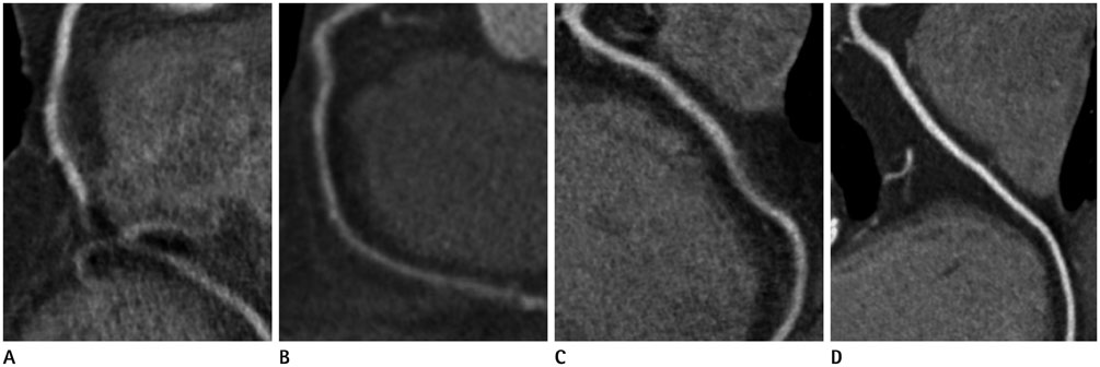
Fig. 1 4-point grading scale of coronary artery image quality. The scores of grade 2 to 4 were regarded diagnostic and grade 1 was non-diagnostic. A. Grade 1 (poor), severe degree of image degradation or discontinuation of vessel contour that prevented vessel lumen evaluation. B. Grade 2 (adequate), moderate degree of image degradation with some obstacle to vessel lumen evaluation. C. Grade 3 (good), minor degree of image degradation without affecting vessel lumen evaluation grade. D. Grade 4 (excellent), no image degradation.
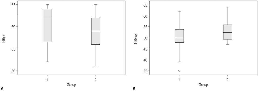
Fig. 2 HRpre and HRmean in group 1 and 2. Boxplots show HRpre (A) and HRmean (B) in subjects administered a low dose β-blocker (group 1) and those that were not (group 2). (A) Mean heart rates (HR) prior to administration of β-blocker (HRpre) are not statistically different between group 1 and group 2 (p = 0.08). HR change (HRchg) was significantly larger in group 1 than in group 2 (p < 0.001) and (B) mean HR during coronary CT angiography (HRmean) in group 1 was significantly lower than group 2 (p = 0.016). Note.-Box = 1st-3rd quartiles, Bold line = median, Whiskers = minimum and maximum values, o = outlier, HR change (HRchg) = difference between HRpre and HRmean (i.e., HRchg = HRpre - HRmean)

Fig. 3 Heart rate variability (HRV) in group 1 and 2. Boxplots show HRV in subjects with low dose β-blocker (group 1) and without β-blocker (group 2). During coronary CT angiography scanning, HRV was not different in group 1 and group 2 (p = 0.147). Note.-Box = 1st-3rd quartiles, Bold line = median, Whiskers = minimum and maximum values, o = outlier

Fig. 4 Linear regression plots of mean image quality scores over the mean heart rate and heart rate variability (HRV) in 75 patients. A. The mean image quality score of the coronary artery segments in both groups is negatively correlated with mean heart rate (r = -0.449, p < 0.001). B. The mean image quality scores for all coronary artery segments and HRV shows no significant correlation (r = -0.013, p = 0.913). Dotted lines represent 95% confidence limits.
Reference
-
1. Raff GL, Gallagher MJ, O'Neill WW, Goldstein JA. Diagnostic accuracy of noninvasive coronary angiography using 64-slice spiral computed tomography. J Am Coll Cardiol. 2005; 46:552–557.2. Mollet NR, Cademartiri F, van Mieghem CA, Runza G, McFadden EP, Baks T, et al. High-resolution spiral computed tomography coronary angiography in patients referred for diagnostic conventional coronary angiography. Circulation. 2005; 112:2318–2323.3. Martuscelli E, Romagnoli A, D'Eliseo A, Razzini C, Tomassini M, Sperandio M, et al. Accuracy of thin-slice computed tomography in the detection of coronary stenoses. Eur Heart J. 2004; 25:1043–1048.4. Leschka S, Scheffel H, Husmann L, Gämperli O, Marincek B, Kaufmann PA, et al. Effect of decrease in heart rate variability on the diagnostic accuracy of 64-MDCT coronary angiography. AJR Am J Roentgenol. 2008; 190:1583–1590.5. Pannu HK, Alvarez W Jr, Fishman EK. Beta-blockers for cardiac CT: a primer for the radiologist. AJR Am J Roentgenol. 2006; 186:6 Suppl 2. S341–S345.6. Leschka S, Wildermuth S, Boehm T, Desbiolles L, Husmann L, Plass A, et al. Noninvasive coronary angiography with 64-section CT: effect of average heart rate and heart rate variability on image quality. Radiology. 2006; 241:378–385.7. Brodoefel H, Reimann A, Burgstahler C, Schumacher F, Herberts T, Tsiflikas I, et al. Noninvasive coronary angiography using 64-slice spiral computed tomography in an unselected patient collective: effect of heart rate, heart rate variability and coronary calcifications on image quality and diagnostic accuracy. Eur J Radiol. 2008; 66:134–141.8. Austen WG, Edwards JE, Frye RL, Gensini GG, Gott VL, Griffith LS, et al. A reporting system on patients evaluated for coronary artery disease. Report of the Ad Hoc Committee for Grading of Coronary Artery Disease, Council on Cardiovascular Surgery, American Heart Association. Circulation. 1975; 51:4 Suppl. 5–40.9. Shuman WP, Branch KR, May JM, Mitsumori LM, Lockhart DW, Dubinsky TJ, et al. Prospective versus retrospective ECG gating for 64-detector CT of the coronary arteries: comparison of image quality and patient radiation dose. Radiology. 2008; 248:431–437.10. Brodoefel H, Burgstahler C, Tsiflikas I, Reimann A, Schroeder S, Claussen CD, et al. Dual-source CT: effect of heart rate, heart rate variability, and calcification on image quality and diagnostic accuracy. Radiology. 2008; 247:346–355.11. Husmann L, Valenta I, Gaemperli O, Adda O, Treyer V, Wyss CA, et al. Feasibility of low-dose coronary CT angiography: first experience with prospective ECG-gating. Eur Heart J. 2008; 29:191–197.12. Scheffel H, Alkadhi H, Leschka S, Plass A, Desbiolles L, Guber I, et al. Low-dose CT coronary angiography in the step-and-shoot mode: diagnostic performance. Heart. 2008; 94:1132–1137.13. Earls JP, Berman EL, Urban BA, Curry CA, Lane JL, Jennings RS, et al. Prospectively gated transverse coronary CT angiography versus retrospectively gated helical technique: improved image quality and reduced radiation dose. Radiology. 2008; 246:742–753.14. Jeong DW, Choo KS, Baik SK, Kim YW, Jeon UB, Kim JS, et al. Step-and-shoot prospectively ECG-gated versus retrospectively ECG-gated with tube current modulation coronary CT angiography using the 128-slice MDCT: comparison of image quality and radiation dose. Acta Radiol. 2011; 52:155–160.15. College of Physicians and College of Radiologists Joint Writing Committee. Thye HK, Han KB, Chen K, Chua K, Kwok R, et al. Guidelines on cardiac CT in Singapore (2006). Ann Acad Med Singapore. 2006; 35:287–296.16. Hanly PJ, George CF, Millar TW, Kryger MH. Heart rate response to breath-hold, valsalva and Mueller maneuvers in obstructive sleep apnea. Chest. 1989; 95:735–739.17. Zhang SZ, Hu XH, Zhang QW, Huang WX. Evaluation of computed tomography coronary angiography in patients with a high heart rate using 16-slice spiral computed tomography with 0.37-s gantry rotation time. Eur Radiol. 2005; 15:1105–1110.18. Lu B, Mao SS, Zhuang N, Bakhsheshi H, Yamamoto H, Takasu J, et al. Coronary artery motion during the cardiac cycle and optimal ECG triggering for coronary artery imaging. Invest Radiol. 2001; 36:250–256.19. Earls JP. How to use a prospective gated technique for cardiac CT. J Cardiovasc Comput Tomogr. 2009; 3:45–51.20. Herzog BA, Husmann L, Burkhard N, Valenta I, Gaemperli O, Tatsugami F, et al. Low-dose CT coronary angiography using prospective ECG-triggering: impact of mean heart rate and heart rate variability on image quality. Acad Radiol. 2009; 16:15–21.21. Hong YJ, Kim SJ, Lee SM, Min PK, Yoon YW, Lee BK, et al. Low-dose coronary computed tomography angiography using prospective ECG-triggering compared to invasive coronary angiography. Int J Cardiovasc Imaging. 2011; 27:425–431.
- Full Text Links
-
- Actions
-
Cited
- CITED
-
- Close
- Share
-
- Similar articles
-
- Technical Aspect of Coronary CT Angiography: Imaging Tips and Safety Issues
- Prospectively Electrocardiogram-Gated High-Pitch Spiral Acquisition Mode Dual-Source CT Coronary Angiography in Patients with High Heart Rates: Comparison with Retrospective Electrocardiogram-Gated Spiral Acquisition Mode
- Coronary CT Angiography
- The Effects of Lidocaine and Propranolol on Heart Rate and Blood Pressure of the Ketamine
- Effect of Healing Beats Program on Stress, Heart Rate and Sleep Quality of Colorectal Cancer Patients Treated with Chemotherapy: A Randomized Controlled Trial
