J Korean Acad Prosthodont.
2014 Apr;52(2):90-96. 10.4047/jkap.2014.52.2.90.
The effect of heat and press-on-metal technique on marginal fit of metal-ceramic crown
- Affiliations
-
- 1Department of Prosthodontics, School of Dentistry, Kyungpook National University, Daegu, Republic of Korea. chlee@knu.ac.kr
- 2Department of Prosthodontics, College of Dentistry, Wonkwang University, Iksan, Republic of Korea.
- KMID: 2000134
- DOI: http://doi.org/10.4047/jkap.2014.52.2.90
Abstract
- PURPOSE
The purpose of this study is to see what impact the heat and press-on-metal technique has on the marginal fit of metal ceramic crown.
MATERIALS AND METHODS
Prior to the experiment, 4 metal master models were prepared. Each model has margin of chamfer, margin of heavy chamfer, margin of shoulder with bevel and margin of shoulder (collarless). Additionally, 10 crowns were made for each margin, total of 40 crowns. Marginal discrepancy between the master model and crown was observed at x100 microscopic magnification in two states; in coping state and upon completion of making metal ceramic crown. Data analysis was performed using paired t-test along with one-way ANOVA and Duncan multiple comparison test.
RESULTS
After analyzing mean and standard deviation of marginal discrepancy, it was confirmed that marginal discrepancies were within the clinical permitted range for all states; in coping state and upon completion of making metal ceramic crown. For the chamfer group, a significant increase in marginal discrepancy upon completion of making metal ceramic crown was observed compared to the heavy chamfer group. Also, a marginal discrepancy of porcelain margin in shoulder group was significantly less than the marginal discrepancy of metal margin in chamfer and shoulder group.
CONCLUSION
From the test result, one can conclude that marginal fit of metal ceramic crown built with heat and press-on-metal technique is not significantly different from marginal fit of metal ceramic crown built with traditional technique. And along with efficiency of this system, heat and press-on-metal technique is considered in clinic.
MeSH Terms
Figure
-

Fig. 1. Four master models used in this study (From the left side, chamfer, heavy chamfer, shoulder with bevel, and shoulder).

Fig. 2. Microscopic measuring of marginal discrepancy at the middle point of labial margin of heavy chamfer. (A) Metal coping, (B) Metal ceramic crown.
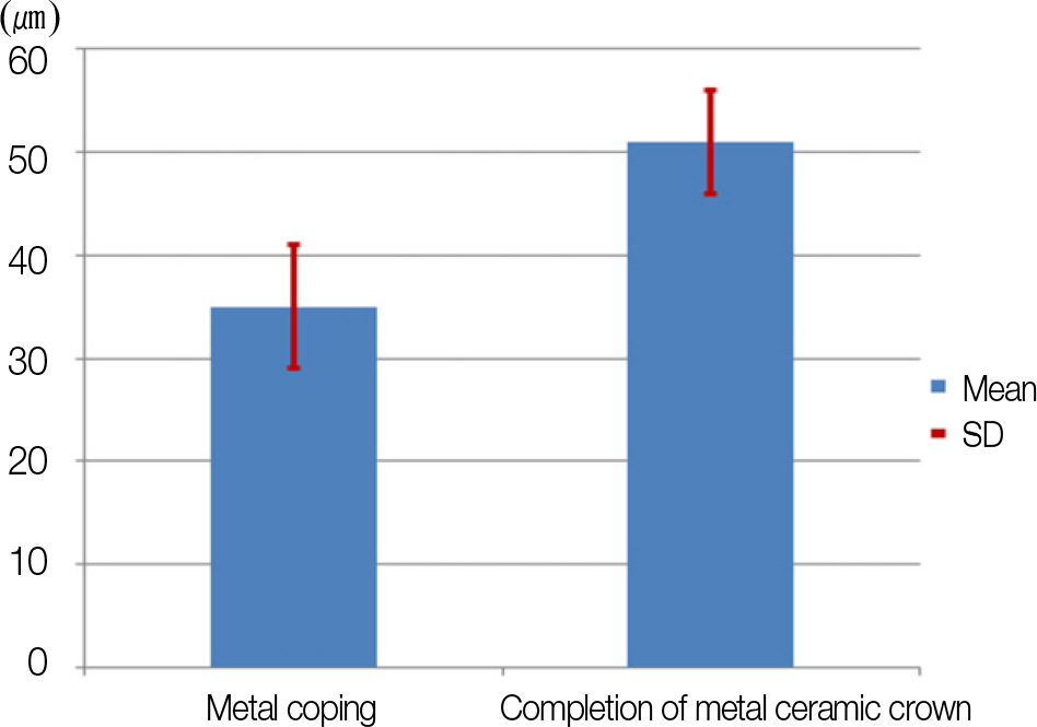
Fig. 3. Comparison of the marginal discrepancy in Group C (㎛). ∗ P-value is determined by t-test procedure.
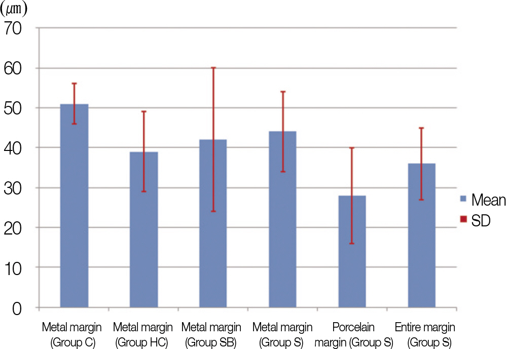
Fig. 4. Comparison of the marginal discrepancies after completion of metal ceramic crown (㎛). ∗ P-value is determined one-way ANOVA procedure.
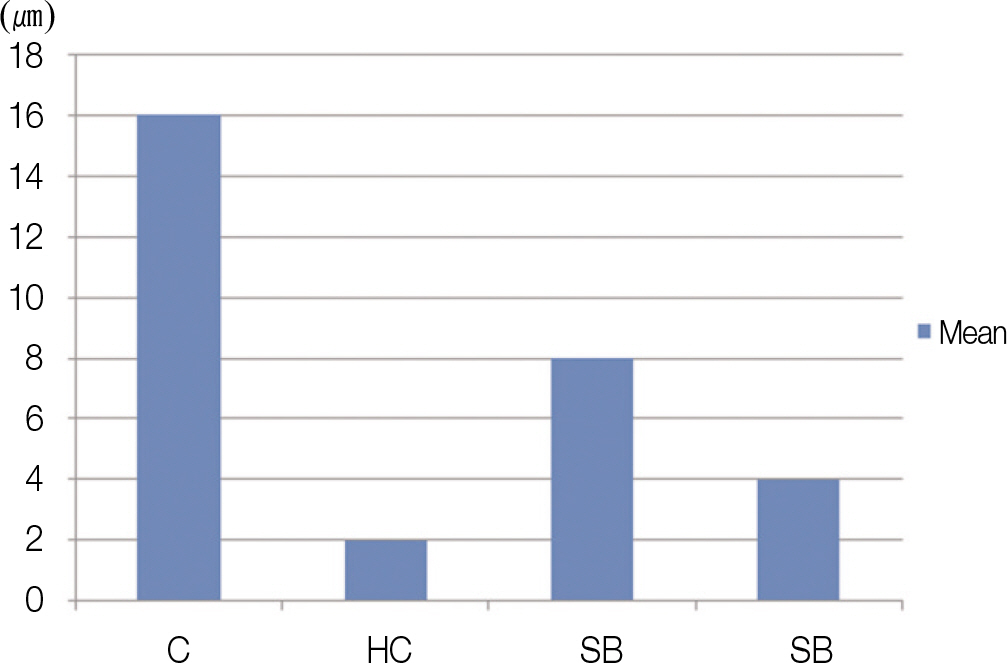
Fig. 5. Comparison between each group in the marginal adaptation discrepancy at the same point of metal margin after completion of metal ceramic crown (㎛). ∗P-value is determined one-way ANOVA procedure.
Reference
-
1.Kim YH., Lee SH. Fitness of the collarless metal-ceramic restorations at labial margins: A scanning electron microscope study. J Korean Acad Prosthodont. 1985. 23:113–24.2.Kim KH. A study of marginal fitness on metal-ceramic restoration. J Korean Dent Assoc. 1985. 23:593–602.3.Yoon IJ., Chang WS., Yang JH., Lee SH. A scanning electron microscopic study on the labial marginal fit of metal ceramic crowns made by different techniques. J Korean Acad Prosthodont. 1986. 24:151–64.4.Belser UC., MacEntee MI., Richter WA. Fit of three porcelain-fused-to-metal marginal designs in vivo: a scanning electron microscope study. J Prosthet Dent. 1985. 53:24–9.
Article5.Shillingburg HT Jr., Hobo S., Fisher DW. Preparation design and margin distortion in porcelain-fused-to-metal restorations. J Prosthet Dent. 1973. 29:276–84.
Article6.Van Rensburg F., Strating H. Evaluation of the marginal integrity of ceramometal restorations: Part II. J Prosthet Dent. 1984. 52:210–4.
Article7.Lee SH. Marginal fit of collarless metal-ceramic crowns: scanning electron microscopy study. J Korean Dent Assoc. 1985. 23:585–91.8.Faucher RR., Nicholls JI. Distortion related to margin design in porcelain-fused-to-metal restorations. J Prosthet Dent. 1980. 43:149–55.
Article9.Hung SH., Hung KS., Eick JD., Chappell RP. Marginal fit of porcelain-fused-to-metal and two types of ceramic crown. J Prosthet Dent. 1990. 63:26–31.
Article10.Kim JH., Yang JH., Lee SH. A comparative study on the marginal fit between castable ceramic(Dicor) crowns and Metal-ceramic crowns. J Kroean Acad Prosthodont. 1988. 26:51–62.11.Gemalmaz D., Alkumru HN. Marginal fit changes during porcelain firing cycles. J Prosthet Dent. 1995. 73:49–54.12.Strating H., Pameijer CH., Gildenhuys RR. Evaluation of the marginal integrity of ceramometal restorations. Part I. J Prosthet Dent. 1981. 46:59–65.13.Omar R. Scanning electron microscopy of the marginal fit of ceramometal restorations with facially butted porcelain margins. J Prosthet Dent. 1987. 58:13–9.
Article14.Hamaguchi H., Cacciatore A., Tueller VM. Marginal distortion of the porcelain- bonded-to-metal complete crown: an SEM study. J Prosthet Dent. 1982. 47:146–53.15.Shillingburg HT Jr., Jacobi R., Brackett SE. Fundamentals of tooth preparations for cast metal and porcelain restorations. Quintessence Publishing Co., Inc.;1987. p. 259–76.16.McLean JW., von Fraunhofer JA. The estimation of cement film thickness by an in vivo technique. Br Dent J. 1971. 131:107–11.
Article
- Full Text Links
-
- Actions
-
Cited
- CITED
-
- Close
- Share
-
- Similar articles
-
- Fabrication of a metal-ceramic crown to fit an existing partial removable dental prosthesis using ceramic pressed to metal technique: a clinical report
- Marginal fit of the digident CAD/CAM zirconia ceramic crowns
- Marginal fit of the auro galvano crown system made using the electroforming technique
- In vitro marginal fit of the computer-aided milled cercon crowns
- Comparative study in marginal fit of a pressed ceramic and feldspathic porcelain fused to metal restoration
