A rare case of primary ovarian leiomyoma with atypical Meigs' syndrome
- Affiliations
-
- 1Department of Obstetrics and Gynecology, Samsung Medical Center, Sungkyunkwan University School of Medicine, Seoul, Korea. garden.lee@samsung.com
- KMID: 1992628
- DOI: http://doi.org/10.5468/KJOG.2012.55.4.285
Abstract
- Primary ovarian leiomyoma is a rare benign ovarian tumor. Ovarian leiomyoma accompanied with atypical Meigs' syndrome is extremely rare. A 13-year-old woman had underlying primary lymphedema and visited our clinic due to associated vulvar edema. During a gynecologic examination, we detected a right ovarian tumor with large amounts of ascites on transrectal ultrasonography. The tumor size increased from 4 to 7 cm during 6 months follow-up. After laparoscopic right ovarian tumorectomy, the final pathology of the tumor was primary ovarian leiomyoma. Ovarian leiomyomas are not typically suspected before surgery due to their extreme rarity and because they are easily misdiagnosed as fibroma during frozen biopsy due to similarities between both diseases. Here we introduce the case of a primary ovarian leiomyoma accompanied with atypical Meigs' syndrome.
Keyword
MeSH Terms
Figure
-
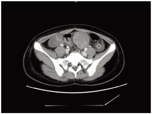
Fig. 1 Poor delineation of the right ovarian mass measuring about 8cm in length on computed tomography.
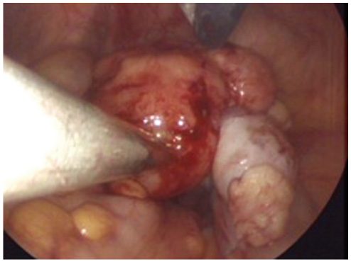
Fig. 2 Gross picture of 5cm sized solid tumors originating from right ovary. The ovarian tumor and the surrounding whitish fragile tissues are separated from right tube and is limited to the right ovary.
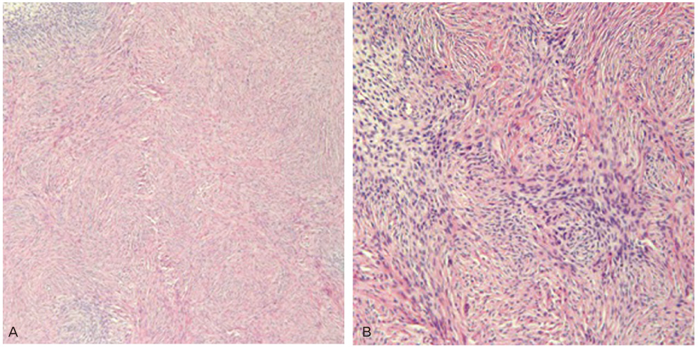
Fig. 3 (A) Spindle-shaped cells with wavy nuclei and extensive hyalinization was seen in the right ovarian tumor (H&E, ×100). (B) On the high powered magnititude, no significant nuclear atypia or pleomorphism was seen (H&E, ×200).
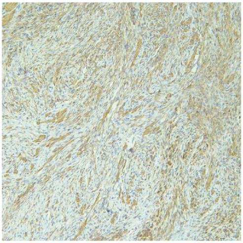
Fig. 4 Immunohistochemically, the tumor cells were strongly positive for smooth muscle actin (smooth muscle actin, ×200).

Fig. 5 The tumor cells were strongly positive for reticulin and the tumor was confirmed as leiomyoma (reticulin, ×200).
Reference
-
1. van Esch EM, van Wijngaarden SE, Schaafsma HE, Smeets MJ, Rhemrev JP. The diagnostic and therapeutic approach of a primary bilateral leiomyoma of the ovaries: a case report and a literature review. Arch Gynecol Obstet. 2011. 283:1369–1371.2. Güney M, Ozsoy M, Oral B, Mungan T, Kapucuoğlu N. Unilateral primary ovarian leiomyoma in adolescent: a case report. Arch Gynecol Obstet. 2007. 275:507–510.3. Koo YJ, Cho YJ, Kim JY, Lee JE, Kim ML, Kim JM, et al. Ovarian leiomyoma as a potential cause of compromised fertility. Fertil Steril. 2011. 95:1120.4. Abdel-Gadir A, Francis ND, Oyawoye OO, Chander BP. Secondary amenorrhoea with high inhibin B level caused by parasitic ovarian leiomyoma. Gynecol Endocrinol. 2010. 26:93–95.5. Kurai M, Shiozawa T, Noguchi H, Konishi I. Leiomyoma of the ovary presenting with Meigs' syndrome. J Obstet Gynaecol Res. 2005. 31:257–262.6. Bucella D, Limbosch JF, Buxant F, Simon P, Fayt I, Anaf V, et al. Recurrence of mitotically active cellular fibroma of the ovary. Obstet Gynecol Int. 2009. 2009:803062.7. Tomas D, Lenicek T, Tuckar N, Puljiz Z, Ledinsky M, Kruslin B. Primary ovarian leiomyoma associated with endometriotic cyst presenting with symptoms of acute appendicitis: a case report. Diagn Pathol. 2009. 4:25.8. Paladini D, Testa A, Van Holsbeke C, Mancari R, Timmerman D, Valentin L. Imaging in gynecological disease (5): clinical and ultrasound characteristics in fibroma and fibrothecoma of the ovary. Ultrasound Obstet Gynecol. 2009. 34:188–195.9. Shiau CS, Chang MY, Hsieh CC, Hsieh TT, Chiang CH. Meigs' syndrome in a young woman with a normal serum CA-125 level. Chang Gung Med J. 2005. 28:587–591.10. Son CE, Choi JS, Lee JH, Jeon SW, Hong JH, Bae JW. Laparoscopic surgical management and clinical characteristics of ovarian fibromas. JSLS. 2011. 15:16–20.
