J Korean Acad Conserv Dent.
2005 Mar;30(2):112-120. 10.5395/JKACD.2005.30.2.112.
Microleakage of the class V cavity according to restoration site and cavity size using SEM and three-dimensional reconstruction techniques
- Affiliations
-
- 1Department of Conservative Dentistry, School of Dentistry, Dankook University, Korea. donyshin@dankook.ac.kr
- KMID: 1987003
- DOI: http://doi.org/10.5395/JKACD.2005.30.2.112
Abstract
- This study was done to evaluate whether there were any differences in microleakage of class V composite restorations according to restoration site and cavity size. Total sixty-four restorations were made in molar teeth using Esthet-X. Small (2 x 2 x 1.5 mm) and large (4 x 2 x 1.5 mm) restorations were made at the buccal/lingual surface and the proximal surface each. After 1,000 times of thermocycling (5degrees C - 55degrees C), resin replica was made and the percentage of marginal gap to the whole periphery of the restoration was estimated from SEM evaluation. Thermocycled tooth was dye penetrated with 50% silver nitrate solution. After imbedding in an auto-curing resin, it was serially ground with a thickness of 0.25 mm. Volumetric microleakage was estimated after reconstructing three dimensionally. Two-way ANOVA and independent T-test for dye volume, Mann-Whitney U test for the percentage of marginal gap, Spearman's rho test for the relationship between two techniques were used. The results were as follows: 1. The site and size of the restoration affected on the microleakage of restoration. Namely, much more leakage was seen in the proximal and the large restorations rather than the buccal/lingual and the small restorations. 2. Close relationship was found between two techniques (Correlation coefficient = 0.614 / P = 0.000). Within the limits of this study, it was noted that proximal and the large restorations leaked more than buccal/lingual and the small restorations. Therefore, it should be strictly recommended large exposure of margins should be avoided by reducing unnecessary tooth reduction.
MeSH Terms
Figure
-

Figure 1 Occlusal view of three-dimensionally reconstructed image (Green : restoration, Red : dye)

Figure 2 Calculation of marginal gap: Percentage of marginal gap to cavity perimeter (SEM image ; × 35) (Blue line : cavity perimeter, Red line : area with marginal gap)
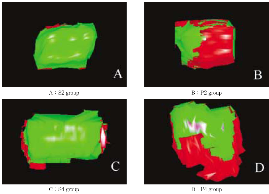
Figure 3 Internal view of three-dimensionally reconstructed images of experimental groups (Green : restoration, Red : dye)
Reference
-
1. Eick JD, Welch FH. Polymerization shrinkage of posterior composite resins and its possible influence on postoperative sensitivity. Quintessence Int. 1986. 17(2):103–111.2. Tjan AH, Bergh BH, Lidner C. Effect of various incremental techniques on the marginal adaptation of Class II composite resin restorations. J Prosthet Dent. 1992. 67(1):62–66.
Article3. Lutz F, Krejci , Oldenburg TR. Elimination of polymerization stresses at the margins of posterior composite resin restorations: A new restorative technique. Quintessence Int. 1986. 17(12):777–784.4. Uno S, Asmussen E. Marginal adaptation of a restorative resin polymerized at reduced rate. Scand J Dent Res. 1991. 99:440–444.
Article5. Burgess JO, DeGoes M, Walker R, Ripps AH. An evaluation of four light-curing units comparing soft and hard curing. Pract Periodontics Aesthet Dent. 1999. 11(1):125–133.6. Aboushala A, Kugel G, Hurley E. Class II composite resin restorations using glass-ionomer liners: Microleakage studies. J Clin Pediatr Dent. 1996. 21(1):67–71.7. Õilo G. Biodegradation of dental composites/glassionomer cements. Adv Dent Res. 1992. 6:50–54.8. Fortin D, Swift EJ Jr, Denehy GE, Reinhardt JW. Bond strength and microleakage of current dentine adhesives. Dent Mater. 1994. 10(4):253–258.9. Rigsby DF, Retief DH, Bidez MW, Russell CM. Effect of axial load and temperature cycling on microleakage of resin restorations. Am J Dent. 1992. 5(3):155–159.10. Donly KJ, Ellis RK. Glass inserts. A new dimension in restorative dentistry. Am J Dent. 1989. 2(1):21–24.11. Feilzer A, de Gee AJ, Davidson CL. Setting stress in composite resin in relation to configuration of the restoration. J Dent Res. 1987. 66:1636–1639.
Article12. Hobson RS, Rugg-Gunn AJ, Booth TA. Acid-etch patterns on the buccal surface of human permanent teeth. Arch Oral Biol. 2002. 47(5):407–412.
Article13. Sturdevant JR, Pashley DH. Regional dentin permeability of Class I and II cavity preparations. J Dent Res. 1989. 68:203. (abstract No. 173).14. Saboia Vde P, Pimenta LA, Ambrosano GM. Effect of collagen removal on microleakage of resin composite restorations. Oper Dent. 2002. 27(1):38–43.15. Adamo HL, Buruiana R, Schertzer L, Boylan RJ. A comparison of MTA, Super-EBA, composite and amalgam as root end filling materials using a bacterial microleakage model. Int Endod J. 1999. 32:197–203.
Article16. Hembree JH, Andrew JT. Microleakage of several class V anterior restorative materials: a laboratory study. J Am Dent Assoc. 1978. 97(2):179–183.
Article17. Manhart J, Chen HY, Mehl A, Weber K, Hickel R. Marginal quality and microleakage of adhesive class V restorations. J Dent. 2001. 29(2):123–130.
Article18. Youngson CC, Jones JC, Fox K, Smith IS, Wood DJ, Gale M. A fluid filtration and clearing technique to assess microleakage associated with three dentine bonding systems. J Dent. 1999. 27(3):223–233.
Article19. Von Fraunhofer JA, Adachi EI, Barnes DM, Romberg E. The effect of tooth preparation on microleakage behavior. Oper Dent. 2000. 25(6):526–533.20. Ha SY, Shin DH. New quantitative measuring technique for microleakage of the restored tooth through 3D reconstruction. J Korean Acad Conserv Dent. 2004. 29(5):413–422.
Article21. Estafan D, Pines MS, Erakin C, Fuerst PF. Microleakage of Class V restorations using two different compomer systems: an in vitro study. J Clin Dent. 1999. 10(4):124–126.22. Hatibovic-Kofman S, Wright GZ, Braverman I. Microleakage of sealants after conventional, bur, and air-abrasion preparation of pits and fissures. Pediatr Dent. 1998. 20(3):173–176.23. Grande RH, Ballester R, Singer Jda M, Santos JF. Microleakage of a universal adhesive used as a fissure sealant. Am J Dent. 1998. 11(3):109–113.24. Mixson J, Eick JD, Chappell RP, Tira DE, Moore DL. Comparison of two-surface and multi-surface scoring methodologies for in vitro microleakage studies. Dent Mater. 1991. 7(3):191–196.
Article25. Shin DH. Dentinal microleakage study on the light curable restorative glass ionomer cement. J Korean Acad Conserv Dent. 1995. 20(2):832–838.26. Arnold WH, Gaengler P, Kalkutschke L. Three-dimensional reconstruction of approximal subsurface caries lesions in deciduous molars. Clin Oral Investig. 1998. 2(4):174–179.
Article27. Mikrogeorgis G, Lyroudia KL, Nikopoulos N, Pitas I, Molyvdas I, Lambrianidis TH. 3D computer-aided reconstruction of six teeth with morphological abnormalities. Int Endod J. 1999. 32(2):88–93.
Article28. Jacobs R, Adriansens A, Verstreken K, Suetens P, van Steenberghe D. Predictability of a three-dimensional planning system for oral implant surgery. Dentomaxillofac Radiol. 1999. 28(2):105–111.
Article29. Jung EH, Shin DH. Morphological analysis of Cshaped root using 3D reconstruction. J Korean Acad Conserv Dent. 2002. 27(4):421–431.
Article30. Veis A, Lambrianides T, Nicolaou A. Area-metric analysis of dye leakage for evaluation of sealing ability of root canal obturation techniques. Endod Dent Traumatol. 1996. 12(5):222–226.
Article31. Gale MS, Darvell BW, Cheung GS. Three-dimensional reconstruction of microleakage pattern using a sequential grinding technique. J Dent. 1994. 22(6):370–375.
Article32. Dorminey JC, Dunn WJ, Taloumis LJ. Shear bond strength of orthodontic brackets bonded with a modified 1-step etchant-and-primer technique. Am J Orthod Dentofacial Orthop. 2003. 124(4):410–413.
Article33. Carvalho RM, Pereira JC, Yoshiyama M, Pashley DH. A review of polymerization contraction: The influence of stress development versus stress relief. Oper Dent. 1996. 21:17–24.34. Asmussen E, Munksgaard EC. Bonding of restorative resins to dentine: Status of dentin adhesives and impact on cavity design and filling techniques. Int Dent J. 1988. 38:97–104.35. Marshall GW Jr, Chang YJ, Gansky SA, Marshall SJ. Demineralization of caries-affected transparent dentin by citric acid: an atomic force microscopy study. Dent Mater. 2001. 17(1):45–52.
Article36. Saunders WP, Saunders EM. Microleakage of bonding agents with wet and dry bonding techniques. Am J Dent. 1996. 9(1):34–36.37. Walls AW, Lee J, McCabe JF. The bonding of composite resin to moist enamel. Br Dent J. 2001. 191(3):148–150.
Article38. Youngson CC, Jones JC, Manogue M, Smith IS. In vitro dentinal penetration by tracers used in microleakage studies. Int Endod J. 1998. 31(2):90–99.
Article39. Hakimeh S, Vaidyanathan J, Houpt ML, Vaidyanathan TK, Von Hagen S. effect of load cycling, thermal cycling, and cavity shape differences. J Prosthet Dent. 2000. 83:194–203.40. Crim GA. Marginal leakage of visible light-cured glass ionomer restorative materials. J Prosthet Dent. 1993. 69:561–563.
Article41. Hilton TJ, Ferracane JL. Cavity preparation factors and microleakage of Class II composite restorations filled at intraoral temperatures. Am J Dent. 1999. 12:123–130.42. Santini A, Mitchell S. Microleakage of composite restorations bonded with three new dentin bonding agents. J Esthet Dent. 1998. 10(6):296–304.
Article
- Full Text Links
-
- Actions
-
Cited
- CITED
-
- Close
- Share
-
- Similar articles
-
- Estimation of relation between techniques of dye penetration for microleakage and SEM evaluation for marginal adaptation of the restoration
- New quantitative measuring technique for microleakage of the restored tooth through 3D reconstruction
- Comparison of microleakage after load cycling for nanofilled composite resin fillings with or without flowable resin lining
- The effect of restorative materials on the stress distribution of class V composite resin restorations: a 3D finite element investigation
- A quantitative analysis about microleakage of all-in-one adhesives
