J Korean Acad Conserv Dent.
2006 Mar;31(2):119-124. 10.5395/JKACD.2006.31.2.119.
The effect of MTAD on the apical leakage of obturated root canals: an electrochemical study
- Affiliations
-
- 1Department of Conservative Dentistry, Samsung Medical Center, Sungkyunkwan University School of Medicine, Korea. dsparkh@smc.samsung.co.kr
- KMID: 1986857
- DOI: http://doi.org/10.5395/JKACD.2006.31.2.119
Abstract
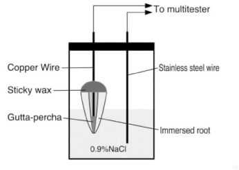
- The purpose of this study was to evaluate the effect of newly developed endodontic root canal cleanser (MTAD) on the apical leakage of obturated root canal using an electrochemical method. Canals of 60 extracted single-rooted human teeth were prepared by using a crown-down technique with rotary nickel-titanium files. In Group 1 (positive control group) and 2 (negative control group), 5.25% NaOCl was used as a canal irrigant and no canal wall treatment was done. In group 3, only 5.25% NaOCl were used as canal irrigant, canal wall treatment and final rinse. In group 4, specimens were irrigated with 5.25% NaOCl, treated with 5 ml of 17% EDTA for 5 minutes and final rinsed with 5.25% NaOCl. Specimens of group 5 were irrigated with 1.3% NaOCl and treated with 5 ml of MTAD for 5 minutes. All root canals are dried with paper points and obtuated with gutta-percha and AH plus as a sealer using a continuous wave of condensation technique except in the group 1. The electrical resistance between the standard and experimental electrodes in canals was measured over a period of 10 days. Rising of apical leakage with time was observed for all the groups. Group 4 and 5 showed lower apical leakage than group 3 but differences between the group 3, 4 and 5 were no statistical significance at any measurement time.
MeSH Terms
Figure
Cited by 1 articles
-
Effect of soft chelating irrigation on the sealing ability of GP/AH Plus root fillings
Yi-Suk Yu, Tae-Gun Kim, Kwang-Won Lee, Mi-Kyung Yu
J Korean Acad Conserv Dent. 2009;34(6):484-490. doi: 10.5395/JKACD.2009.34.6.484.
Reference
-
1. McComb D, Smith DC. A preliminary scanning electron microscopic study of root canals after endodontic procedures. J Endod. 1975. 1(7):238–242.
Article2. Torabinejad M, Handysides R, Khademi A, Bakland LK. Clinical implications of the smear layer in endodontics: a review. Oral Surg Oral Med Oral Pathol Oral Radiol Endod. 2002. 94(6):658–666.
Article3. Baumgartner JC, Mader CL. A scanning electron microscopic evaluation of four root canal irrigation regimens. J Endod. 1987. 13(4):147–157.
Article4. Yamada RS, Armas A, Goldman M, Lin PS. A scanning electron microscopic comparison of a high volume flush with several irrigating solutions. Part 3. J Endod. 1983. 9(4):137–142.
Article5. Gettleman BH, Messer HH, ElDeeb ME. Adhesion of sealer cements to dentin with and without the smear layer. J Endod. 1991. 17(1):15–20.
Article6. Economides N, Liolios E, Kolokuris I, Beltes P. Long-term evaluation of the influence of smear layer removal on the sealing ability of different sealers. J Endod. 1999. 25(2):123–125.
Article7. Kennedy WA, Walker WA, Gough RW. Smear layer removal effects on apical leakage. J Endod. 1986. 12(1):21–27.
Article8. Cergneux M, Ciucchi B, Dietschi JM, Holz J. The influence of the smear layer on the sealing ability of canal obturation. Int Endod J. 1987. 20(5):228–232.
Article9. Vassiliadis L, Liolios E, Kouvas V, Economides N. Effect of smear layer on coronal microleakage. Oral Surg Oral Med Oral Pathol Oral Radiol Endod. 1996. 82(3):315–320.
Article10. Taylor JK, Jeansonne BG, Lemon RR. Coronal leakage: Effects of smear layer, obturation technique, and sealer. J Endod. 1997. 23(8):508–512.
Article11. Karagöz-Küçükay I, Bayirli G. An apical leakage study in the presence and absence of the smear layer. Int Endod J. 1994. 27(2):87–93.
Article12. Saunders WP, Saunders EM. The effect of smear layer upon the coronal leakage of gutta percha root filling and a glass ionomer sealer. Int Endod J. 1992. 25(5):245–249.
Article13. Evans JT, Simon JHS. Evaluation of the apical seal produced by injected thermoplasticized gutta-percha in the absence of smear layer and root canal sealer. J Endod. 1986. 12(3):100–107.
Article14. Chailertvanitkul P, Saunders WP, Mackenzie D. The effect of smear layer on microbial coronal leakage of gutta-percha root fillings. Int Endod J. 1996. 29(4):242–248.
Article15. Saunders WP, Saunders EM. Influence of smear layer and the coronal leakage of thermafil and laterally condensed gutta-percha root fillings with a glass ionomer sealer. J Endod. 1994. 20(4):155–158.
Article16. Madison S, Krell KV. Comparison of ethylenediamine tetracetic acid and sodium hypochlorite on the apical seal of endodontically treated teeth. J Endod. 1984. 10(10):499–503.
Article17. Timpawat S, Sripanaratanakul S. Apical sealing ability of glass ionomer sealer with and without smear layer. J Endod. 1998. 24(5):343–345.
Article18. Timpawat S, Vongsavan N, Messer HH. Effect of removal of the smear layer on apical microleakage. J Endod. 2001. 27(5):351–353.
Article19. Torabinejad M, Khademi AA, Babagoli J, Cho Y, Johnson WB, Bozhilov K, Kim J, Shabahang S. A new solution for the removal of the smear layer. J Endod. 2003. 29(3):170–175.
Article20. Park DS, Lee HJ, Yoo HM, Oh TS. Effect of Nd:YAG laser irradiation on the apical leakage of obturated root canals: an electrochemical study. Int Endod J. 2001. 34(4):318–321.
Article21. Baumgartner JC, Brown CM, Mader CL, Peters DD, Shulman JD. A scanning electron microscopic evaluation of root canal debridement using saline, sodium hypochlorite and citric acid. J Endod. 1984. 10(11):525–531.
Article22. Torabinejad M, Cho Y, Khademi AA, Bakland LK, Shabahang S. The effect of various concentrations of sodium hypochlorite on the ability of MTAD to remove smear layer. J Endod. 2003. 29(4):233–239.
Article23. Delivanis PD, Chapman KA. Comaprison and reliability of techniques for measuring leakage and marginal penetrations. Oral Surg. 1982. 53(4):410–416.
Article
- Full Text Links
- Actions
-
Cited
- CITED
-
- Close
- Share
- Similar articles
-
- Effect of two different calcium hydroxide paste removal techniques on apical leakage: an electrochemical study
- The effect of MTAD as a final root canal irrigants on the coronal bacterial leakage of obturated root canals
- An electrochemical study of the sealing ability of three retrofilling materials
- Bacterial leakage and micro-computed tomography evaluation in round-shaped canals obturated with bioceramic cone and sealer using matched single cone technique
- Apical prepration size in infected root canals


