Anatomical Variation of the Glissonean Pedicle of the Right Liver
- Affiliations
-
- 1Department of Surgery, Ajou University School of Medicine, Korea. wanghj@ajou.ac.kr
- KMID: 1980948
- DOI: http://doi.org/10.14701/kjhbps.2011.15.2.101
Abstract
- PURPOSE
Many studies have been conducted to date regarding whether the right hepatic vein is the accurate border that divides the anterior and posterior section of the right liver. It has been reported that the Glisson pedicle of the right liver may be an anatomical variation that does not have a consistent morphology. We analyzed the relationship between the true borders of the anterior and posterior sections, and the right hepatic vein, based on cadaver dissection and MD-CT image analysis of the anatomical variation of the Glisson pedicle of the right liver.
METHODS
Sixteen cadaver livers were available for dissection from the Department of Anatomy, and pre-operative MD-CTs of 20 donor livers who underwent living donor liver transplantation prior to December 2009, were obtained. We analyzed the 3D-relationship between the branches of the Glisson pedicles and the right hepatic vein of the right liver. They were divided into 3 groups according to the sliding pattern of the branches of the Glisson pedicle origin. When all segmental branches of the anterior pedicle arise from the main trunk of the anterior pedicle and all branches of posterior pedicle arise from the main trunk of posterior pedicle, it was designated as Group A (Normal Group). When a portion of the segmental branches of the anterior pedicle arises from the main trunk of the posterior pedicle, it was designated as Group B (Posterior dominant group). When a portion of the branches of the posterior pedicle arises from the main trunk of the anterior pedicle, it was designated as Group C (Anterior dominant group).
RESULTS
Among the 16 cadaver liver dissections, 6 cases were in Group A, 5 in Group B, and 3 in Group C. Two cases were excluded from the study because the inferior right hepatic vein was the main draining vein of the right liver. The analysis of preoperative MD-CT of the 20 donor livers showed that there were 13, 4, and 3 patients in Groups A, B, and C, respectively.
CONCLUSION
According to Couinaud's theory of anatomy, the right hepatic vein serves as the border between the anterior and posterior sections of the right liver. But, due to the frequent anatomical variations, an adequate understanding of the anatomical variations of the right Glisson pedicle should be necessary for liver surgery.
Keyword
MeSH Terms
Figure
-
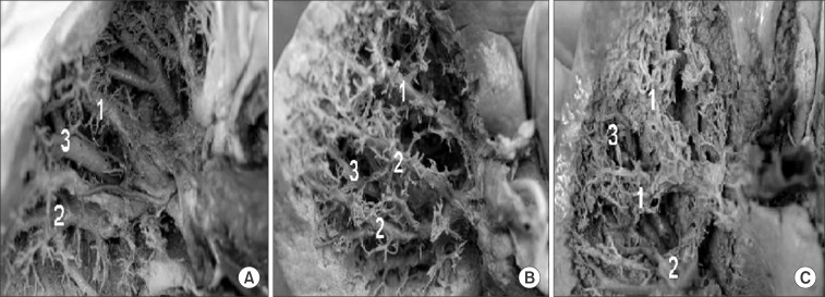
Fig. 1 Dissected pictures of cadaver liver. (A) Normal group: all right posterior branches from the right posterior portal pedicle or all right anterior branches from the right anterior portal pedicle. (B) Right posterior dominant group: some right anterior branches from the right posterior portal pedicle. (C) Right anterior dominant group: some right posterior branches from the anterior portal pedicle. 1: Right anterior portal pedicle, 2: Right posterior portal pedicle, 3: Right hepatic vein.
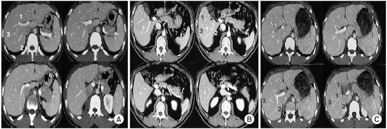
Fig. 2 (A) Normal group: all right posterior branches from the right posterior portal pedicle or all right anterior branches from the right anterior portal pedicle. (B) Right posterior dominant group: some right anterior branches from the right posterior portal pedicle. (C) Right anterior dominant group: some right posterior branches from the anterior portal pedicle. 1: Right anterior portal pedicle, 2: Right posterior portal pedicle, 3: Right hepatic vein.
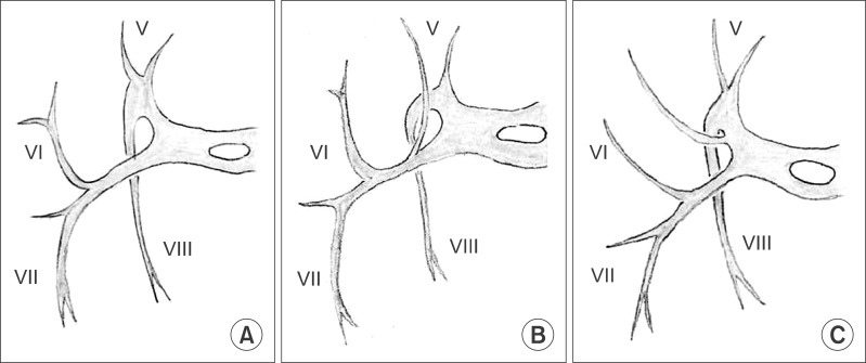
Fig. 3 Variations of the right pedicle. (A) No variations of the right pedicle. (B) Branch of V originating from the right posterior pedicle. (C) Branch of VI originating from the right anterior pedicle (caudal view).
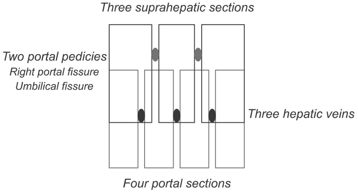
Fig. 4 Diagram of the two segmentations.
Reference
-
1. Couinaud C. Liver anatomy: portal (and suprahepatic) or biliary segmentation. Dig Surg. 1999; 16:459–467. PMID: 10805544.
Article2. Okamoto E, Yamanaka N. Anatomical resection of the right hepatic subsegments preceded by suprahilar ligation of the portal pedicles. In : Tobe T, Kameda H, Okudaira M, editors. Primary liver cancer in Japan. 1992. Tokyo: Springer;p. 229.3. Launois B, Jamieson G. Modern operative techniques in liver surgery. 1993. Edinburgh: Churchill Livingstone.4. Makuuchi M. Knack & pitfalls of liver surgery.(Kr). 2003. 1st ed. Seoul: Koon Ja Publishing co;p. 102–103.5. Belghiti J, Clavien PA, Gadzijev E, et al. The Brisbane 2000 terminology of liver anatomy and resections. HPB. 2000; 2:333–339.
Article6. Cho A, Okazumi S, Miyazawa Y, et al. Proposal for a reclassification of liver based anatomy on portal ramifications. Am J Surg. 2005; 189:195–199. PMID: 15720989.
Article7. Won TW, Park DE, Lee YH, Chae KM. A new classification of the right portal vein using 64 channel multi-dectector CT (MDCT). J Korean Surg Soc. 2008; 75:96–101.
- Full Text Links
-
- Actions
-
Cited
- CITED
-
- Close
- Share
-
- Similar articles
-
- Clinical Application of Hepatic Resection Using Glissonean Pedicle Transection Method and Hanging Maneuver
- Application of temporary inflow control of the Glissonean pedicle method provides a safe and easy technique for totally laparoscopic hemihepatectomy by Glissonean approach
- Laparoscopic drainage basin hepatectomy based on cone unit
- Entry Point of Pedicle Screw in the Lower Lumbar Spine
- Ideal Insertion Point and Angle of Cervical Pedicular Screws in Korean
