The frog appliance for upper molar distalization: a case report
- Affiliations
-
- 1Karadeniz Technical University, Department of Orthodontics, Turkey. dtmehmetbayram@yahoo.com
- KMID: 1975626
- DOI: http://doi.org/10.4041/kjod.2010.40.1.50
Abstract
- The purpose of this article was to evaluate the effects of a new upper molar distalization system, the Frog Appliance, on dentofacial structures in a Class II, division 1 patient. An 11-year-old girl was referred to our clinic for orthodontic treatment. She had a mild skeletal Class II malocclusion with Class II molar and canine relationship on both sides. The treatment plan included distalization of the upper first molars bilaterally followed by full fixed appliance therapy. For the upper molar distalization, a new system, the Frog Appliance, was constructed and applied. Lateral cephalometric radiographs were used to evaluate the treatment results. Distalization of the upper first molars was achieved in four months successfully, and Class I molar relationship was obtained. Total treatment time was 16 months. According to the results of the cephalometric evaluation, a nearly bodily distal molar movement with a slight anchorage loss was attained. In conclusion, the Frog Appliance was found to be a simple, effective, non-invasive, and compliance-free intraoral distalization appliance for achieving bilateral molar distalization.
Keyword
MeSH Terms
Figure
-

Fig 1 Facial and intraoral photographs of the case before treatment (age 11 years).
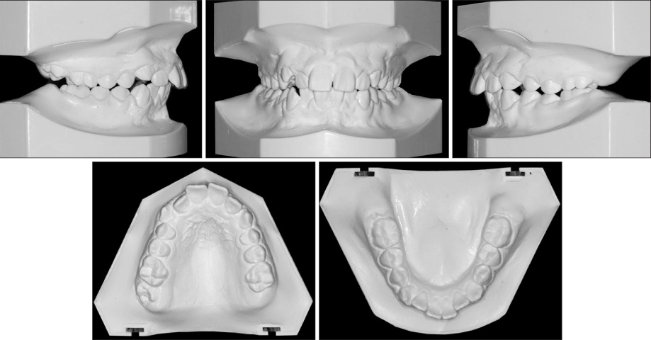
Fig 2 Pretreatment dental casts of the case.

Fig 3 Pretreatment lateral cephalometric and panoramic radiographs of the case.
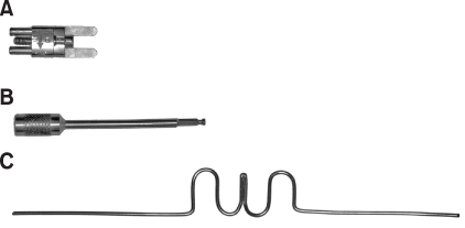
Fig 4 Parts of the Frog Appliance. A, Screw; B, screw driver; C, preformed spring.

Fig 5 Occlusal views of the Frog Appliance. A, During activation; B, on the dental cast and immediately after the cementation (C, D).

Fig 6 Upper occlusal view of the patient immediately after the distalization (A), and intraoral photographs after cutting of the anchor wires of premolars (B-D) (after 4 months of distalization).

Fig 7 Lateral cephalometric and panoramic radiographs of the case taken immediately after the distalization.

Fig 8 Facial and intraoral photographs of the case at the end of the fixed orthodontic treatment (age 12 years 4 months).

Fig 9 Postreatment dental casts of the case.

Fig 10 Postreatment lateral cephalometric and panoramic radiographs of the case.

Fig 11 Local and total superimpositions of the lateral cephalometric tracings before treatment (solid line), after distalization (dotted line) and after treatment (dashed line).

Fig 12 Schematic drawing of distalization effect on the maxillary dentition clearly shows an explicit distal molar movement with a slight anchorage loss on the premolars and the incisors.
Cited by 4 articles
-
Combined treatment with headgear and the Frog appliance for maxillary molar distalization: a randomized controlled trial
Ahmad Sharafeddin Burhan
Korean J Orthod. 2013;43(2):101-109. doi: 10.4041/kjod.2013.43.2.101.Comparison of transverse dental changes induced by the palatally applied Frog appliance and buccally applied Karad's integrated distalizing system
Fatma Deniz Uzuner, Emine Kaygisiz, Fatih Unver, Tuba Tortop
Korean J Orthod. 2016;46(2):96-103. doi: 10.4041/kjod.2016.46.2.96.Cone-beam computed tomography-guided three-dimensional evaluation of treatment effectiveness of the Frog appliance
Mujia Li, Xiaoxia Su, Yang Li, Xianglin Li, Xinqin Si
Korean J Orthod. 2019;49(3):161-169. doi: 10.4041/kjod.2019.49.3.161.Zygoma-gear appliance for intraoral upper molar distalization
Metin Nur, Mehmet Bayram, Alper Pampu
Korean J Orthod. 2010;40(3):195-206. doi: 10.4041/kjod.2010.40.3.195.
Reference
-
1. Gelgör IE, Büyükyilmaz T, Karaman AI, Dolanmaz D, Kalayci A. Intraosseous screw-supported upper molar distalization. Angle Orthod. 2004. 74:838–850.2. Cangialosi TJ, Meistrell ME Jr, Leung MA, Ko JY. A cephalometric appraisal of edgewise Class II nonextraction treatment with extraoral force. Am J Orthod Dentofacial Orthop. 1988. 93:315–324.
Article3. Hilgers JJ. The pendulum appliance for Class II non-compliance therapy. J Clin Orthod. 1992. 26:706–714.4. Ghosh J, Nanda RS. Evaluation of an intraoral maxillary molar distalization technique. Am J Orthod Dentofacial Orthop. 1996. 110:639–646.
Article5. Byloff FK, Darendeliler MA. Distal molar movement using the pendulum appliance. Part 1: clinical and radiological evaluation. Angle Orthod. 1997. 67:249–260.6. Bussick TJ, McNamara JA Jr. Dentoalveolar and skeletal changes associated with the pendulum appliance. Am J Orthod Dentofacial Orthop. 2000. 117:333–343.
Article7. Kinzinger GS, Fritz UB, Sander FG, Diedrich PR. Efficiency of a pendulum appliance for molar distalization related to second and third molar eruption stage. Am J Orthod Dentofacial Orthop. 2004. 125:8–23.
Article8. Gulati S, Kharbanda OP, Parkash H. Dental and skeletal changes after intraoral molar distalization with sectional jig assembly. Am J Orthod Dentofacial Orthop. 1998. 114:319–327.
Article9. Haydar S, Uner O. Comparison of Jones jig molar distalization appliance with extraoral traction. Am J Orthod Dentofacial Orthop. 2000. 117:49–53.
Article10. Brickman CD, Sinha PK, Nanda RS. Evaluation of the Jones jig appliance for distal molar movement. Am J Orthod Dentofacial Orthop. 2000. 118:526–534.
Article11. Carano A, Testa M. The distal jet for upper molar distalization. J Clin Orthod. 1996. 30:374–380.12. Ngantung V, Nanda RS, Bowman SJ. Posttreatment evaluation of the distal jet appliance. Am J Orthod Dentofacial Orthop. 2001. 120:178–185.
Article13. Bolla E, Muratore F, Carano A, Bowman SJ. Evaluation of maxillary molar distalization with the distal jet: a comparison with other contemporary methods. Angle Orthod. 2002. 72:481–494.14. Keles A, Pamukcu B, Tokmak EC. Bilateral maxillary molar distalization with sliding mechanics: keles Slider. World J Orthod. 2002. 3:57–66.15. Fortini A, Lupoli M, Giuntoli F, Franchi L. Dentoskeletal effects induced by rapid molar distalization with the first class appliance. Am J Orthod Dentofacial Orthop. 2004. 125:697–704.
Article16. Cetlin NM, Ten Hoeve A. Nonextraction treatment. J Clin Orthod. 1983. 17:396–413.17. Kim SJ, Chun YS, Jung SH, Park SH. Three dimensional analysis of tooth movement using different types of maxillary molar distalization appliances. Korean J Orthod. 2008. 38:376–387.
Article18. Karaman AI, Basciftci FA, Polat O. Unilateral distal molar movement with an implant-supported distal jet appliance. Angle Orthod. 2002. 72:167–174.19. Keles A, Erverdi N, Sezen S. Bodily distalization of molars with absolute anchorage. Angle Orthod. 2003. 73:471–482.20. Kinzinger GS, Diedrich PR, Bowman SJ. Upper molar distalization with a miniscrew-supported Distal Jet. J Clin Orthod. 2006. 40:672–678.21. Escobar SA, Tellez PA, Moncada CA, Villegas CA, Latorre CM, Oberti G. Distalization of maxillary molars with the bone-supported pendulum: a clinical study. Am J Orthod Dentofacial Orthop. 2007. 131:545–549.
Article
- Full Text Links
-
- Actions
-
Cited
- CITED
-
- Close
- Share
-
- Similar articles
-
- Zygoma-gear appliance for intraoral upper molar distalization
- Comparison of transverse dental changes induced by the palatally applied Frog appliance and buccally applied Karad's integrated distalizing system
- Noncompliance screw supported maxillary molar distalization in a parallel manner
- Combined treatment with headgear and the Frog appliance for maxillary molar distalization: a randomized controlled trial
- C-activator treatment for distalization of maxillary molars in Class II anterior deep bite malocclusion
