Alveolar bone thickness and lower incisor position in skeletal Class I and Class II malocclusions assessed with cone-beam computed tomography
- Affiliations
-
- 1Department of Orthodontics, Faculty of Dentistry, Izmir Katip Celebi University, Izmir, Turkey. baysalasli@hotmail.com
- 2Department of Orthodontics, Faculty of Dentistry, Erciyes University, Kayseri, Turkey.
- 3Department of Orthodontics, Faculty of Dentistry, Adnan Menderes University, Aydin, Turkey.
- KMID: 1975128
- DOI: http://doi.org/10.4041/kjod.2013.43.3.134
Abstract
OBJECTIVE
To evaluate lower incisor position and bony support between patients with Class II average- and high-angle malocclusions and compare with the patients presenting Class I malocclusions.
METHODS
CBCT records of 79 patients were divided into 2 groups according to sagittal jaw relationships: Class I and II. Each group was further divided into average- and high-angle subgroups. Six angular and 6 linear measurements were performed. Independent samples t-test, Kruskal-Wallis, and Dunn post-hoc tests were performed for statistical comparisons.
RESULTS
Labial alveolar bone thickness was significantly higher in Class I group compared to Class II group (p = 0.003). Lingual alveolar bone angle (p = 0.004), lower incisor protrusion (p = 0.007) and proclination (p = 0.046) were greatest in Class II average-angle patients. Spongious bone was thinner (p = 0.016) and root apex was closer to the labial cortex in high-angle subgroups when compared to the Class II average-angle subgroup (p = 0.004).
CONCLUSIONS
Mandibular anterior bony support and lower incisor position were different between average- and high-angle Class II patients. Clinicians should be aware that the range of lower incisor movement in high-angle Class II patients is limited compared to average- angle Class II patients.
MeSH Terms
Figure
-
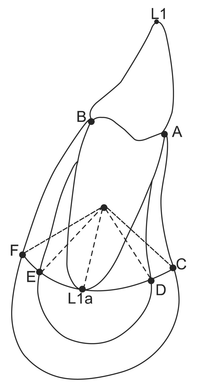
Figure 1 Landmark points at the symphysis and mandibular incisor. L1a, Lower central incisor apex; point A, the most anterior-superior point of mandibular alveolar bone; point B, the most posterior-superior point of the mandibular alveolar bone; point C, the most anterior point of the mandibular alveolar bone; point F, the most posterior point of the mandibular alveolar bone; point D, the inner contour of the anterior cortical plate; point E, the inner contour of the posterior cortical plate (points C, D, E, and F were defined on the trajectory of the hypothetical tipping movement of the mandibular central incisor root around the center of rotation); labial cortical bone thickness (D-C), the length of the arc between points D and C; lingual cortical bone thickness (F-E), the length of the arc between points F and E; alveolar spongious bone thickness (E-D), the length of the arc between points E and D; alveolar spongious and cortical bone thickness (F-C), the length of the arc between points F and C; L1a-D, the length of the arc between points L1a and D; L1a-E, the length of the arc between points L1a and E.
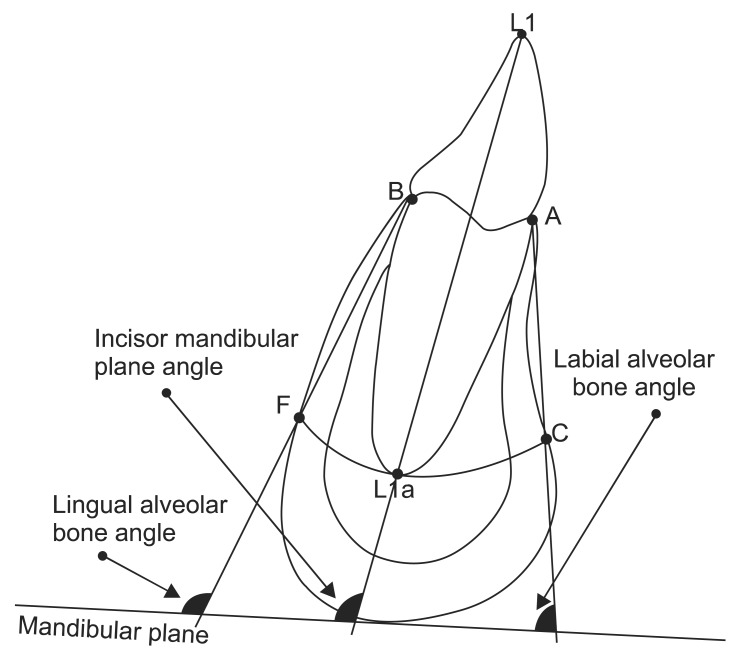
Figure 2 Definition of angular measurements. Abbreviations were given in Figure 1 in detail. The description of the measurements were as follows: Incisor mandibular plane angle, the angle between the central incisor axis and the mandibular plane; labial alveolar bone angle, the angle between line A-C and the mandibular plane; lingual alveolar bone angle; the angle between line B-F and the mandibular line.
Figure 3 Arch length measurements.
Reference
-
1. Mihalik CA. Long term follow-up of Class II adults treated with orthodontic camouflage: a comparison with orthognathic surgery outcomes [thesis]. Chapel Hill: University of North Carolina;2002.2. Proffit WR, Fields HW. Contemporary orthodontics. 3rd ed. St. Louis: Mosby;2000. p. 273.3. Yared KF, Zenobio EG, Pacheco W. Periodontal status of mandibular central incisors after orthodontic proclination in adults. Am J Orthod Dentofacial Orthop. 2006; 130:6.e1–6.e8. PMID: 16849063.
Article4. Ten Hoeve A, Mulie RM. The effect of antero-postero incisor repositioning on the palatal cortex as studied with laminagraphy. J Clin Orthod. 1976; 10:804–822. PMID: 1069732.5. Handelman CS. The anterior alveolus: its importance in limiting orthodontic treatment and its influence on the occurrence of iatrogenic sequelae. Angle Orthod. 1996; 66:95–109. PMID: 8712499.6. Mulie RM, Hoeve AT. The limitations of tooth movement within the symphysis, studied with laminagraphy and standardized occlusal films. J Clin Orthod. 1976; 10:882–893. PMID: 1074868.7. Gracco A, Luca L, Bongiorno MC, Siciliani G. Computed tomography evaluation of mandibular incisor bony support in untreated patients. Am J Orthod Dentofacial Orthop. 2010; 138:179–187. PMID: 20691359.
Article8. Tsunori M, Mashita M, Kasai K. Relationship between facial types and tooth and bone characteristics of the mandible obtained by CT scanning. Angle Orthod. 1998; 68:557–562. PMID: 9851354.9. Kohakura S, Kasai K, Ohno I, Kanazawa E. Relationship between maxillofacial morphology and morphological characteristics of vertical sections of the mandible obtained by CT scanning. J Nihon Univ Sch Dent. 1997; 39:71–77. PMID: 9293703.
Article10. Swasty D, Lee J, Huang JC, Maki K, Gansky SA, Hatcher D, et al. Cross-sectional human mandibular morphology as assessed in vivo by cone-beam computed tomography in patients with different vertical facial dimensions. Am J Orthod Dentofacial Orthop. 2011; 139(4 Suppl):e377–e389. PMID: 21435546.
Article11. Huang J, Bumann A, Mah J. Three-dimensional radiographic analysis in orthodontics. J Clin Orthod. 2005; 39:421–428. PMID: 16100415.12. Yamada C, Kitai N, Kakimoto N, Murakami S, Furukawa S, Takada K. Spatial relationships between the mandibular central incisor and associated alveolar bone in adults with mandibular prognathism. Angle Orthod. 2007; 77:766–772. PMID: 17685772.
Article13. Fuhrmann R. Three-dimensional interpretation of periodontal lesions and remodeling during orthodontic treatment. Part III. J Orofac Orthop. 1996; 57:224–237. PMID: 8765798.14. Fuhrmann RA, Wehrbein H, Langen HJ, Diedrich PR. Assessment of the dentate alveolar process with high resolution computed tomography. Dentomaxillofac Radiol. 1995; 24:50–54. PMID: 8593909.
Article15. Vandenberghe B, Jacobs R, Yang J. Detection of periodontal bone loss using digital intraoral and cone beam computed tomography images: an in vitro assessment of bony and/or infrabony defects. Dentomaxillofac Radiol. 2008; 37:252–260. PMID: 18606746.16. Davidian EJ. Use of a computer model to study the force distribution on the root of the maxillary central incisor. Am J Orthod. 1971; 59:581–588. PMID: 5280425.
Article17. Christiansen RL, Burstone CJ. Centers of rotation within the periodontal space. Am J Orthod. 1969; 55:353–369. PMID: 5251429.
Article18. Ellis E 3rd, McNamara JA Jr. Cephalometric evaluation of incisor position. Angle Orthod. 1986; 56:324–344. PMID: 3466560.19. Garib DG, Yatabe MS, Ozawa TO, Filho OGS. Alveolar bone morphology under the perspective of the computed tomography: Defining the biological limits of tooth movement. Dental Press J Orthod. 2010; 15:192–205.20. Schudy FF. The rotation of the mandible resulting from growth: its implications in orthodontic treatment. Angle Orthod. 1965; 35:36–50. PMID: 14258830.21. Bjork A. Variations in the growth pattern of the human mandible: longitudinal radiographic study by the implant method. J Dent Res. 1963; 42:400–411. PMID: 13971295.
Article22. Nielsen IL. Vertical malocclusions: etiology, development, diagnosis and some aspects of treatment. Angle Orthod. 1991; 61:247–260. PMID: 1763835.
- Full Text Links
-
- Actions
-
Cited
- CITED
-
- Close
- Share
-
- Similar articles
-
- Comparison of interradicular distances and cortical bone thickness in Thai patients with Class I and Class II skeletal patterns using cone-beam computed tomography
- Assessment of lower incisor alveolar bone width using cone-beam computed tomography images in skeletal Class III adults of different vertical patterns
- Crown-root angulations of the maxillary anterior teeth according to malocclusions: A cone-beam computed tomography study in Korean population
- A study on sagittal root position of maxillary anterior teeth in Korean
- Analysis of the root position of the maxillary incisors in the alveolar bone using cone-beam computed tomography
