Accuracy and precision of integumental linear dimensions in a three-dimensional facial imaging system
- Affiliations
-
- 1Department of Orthodontics, School of Dentistry, Kyung Hee University, Seoul, Korea.
- 2Department of Orthodontics, Oral Biology Research Institute, Kyung Hee University, Seoul, Korea. ygpark@khu.ac.kr
- KMID: 1974801
- DOI: http://doi.org/10.4041/kjod.2015.45.3.105
Abstract
OBJECTIVE
A recently developed facial scanning method uses three-dimensional (3D) surface imaging with a light-emitting diode. Such scanning enables surface data to be captured in high-resolution color and at relatively fast speeds. The purpose of this study was to evaluate the accuracy and precision of 3D images obtained using the Morpheus 3D(R) scanner (Morpheus Co., Seoul, Korea).
METHODS
The sample comprised 30 subjects aged 24-34 years (mean 29.0 +/- 2.5 years). To test the correlation between direct and 3D image measurements, 21 landmarks were labeled on the face of each subject. Sixteen direct measurements were obtained twice using digital calipers; the same measurements were then made on two sets of 3D facial images. The mean values of measurements obtained from both methods were compared. To investigate the precision, a comparison was made between two sets of measurements taken with each method.
RESULTS
When comparing the variables from both methods, five of the 16 possible anthropometric variables were found to be significantly different. However, in 12 of the 16 cases, the mean difference was under 1 mm. The average value of the differences for all variables was 0.75 mm. Precision was high in both methods, with error magnitudes under 0.5 mm.
CONCLUSIONS
3D scanning images have high levels of precision and fairly good congruence with traditional anthropometry methods, with mean differences of less than 1 mm. 3D surface imaging using the Morpheus 3D(R) scanner is therefore a clinically acceptable method of recording facial integumental data.
Keyword
MeSH Terms
Figure
-
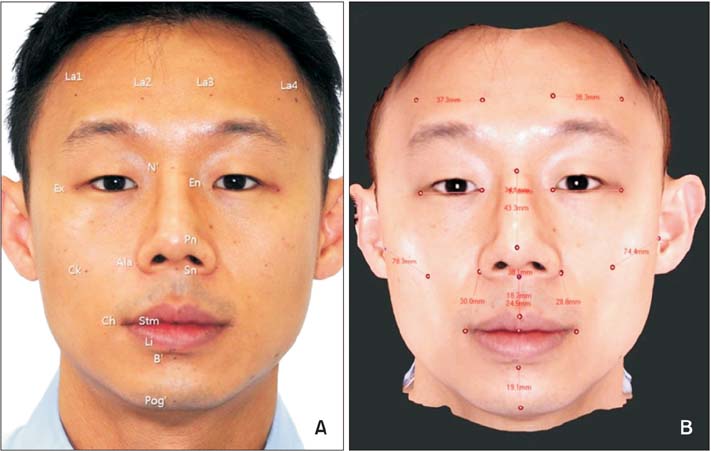
Figure 1 A, Craniofacial landmarks used in this study.11 Tragion (Tra) is not shown. B, Linear distances measured using the Morpheus 3D® scanner. La1-4: points located 5 cm above the right exocanthion, right endocanthion, left endocanthion, and left exocanthion.
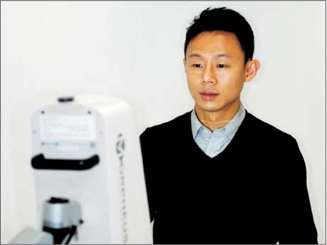
Figure 2 Patients sat with natural head position and reposed lips.
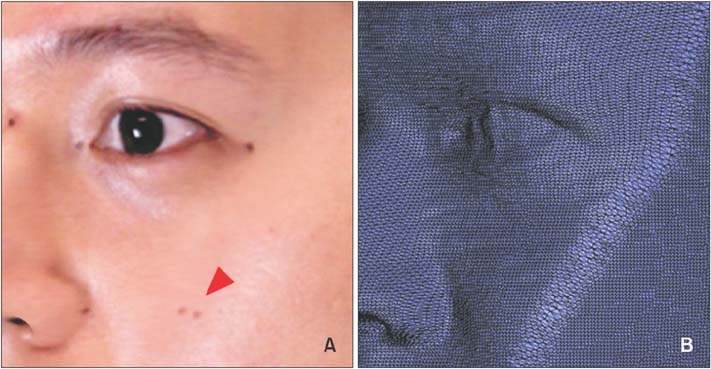
Figure 3 A, Doubled landmark (arrow). B, Integration line, showing its proximity to the doubled landmark.
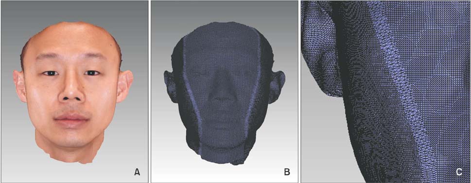
Figure 4 A, A single composite image derived from three images (front, right, and left). B and C, Integration lines in a three-dimensional image.
Cited by 1 articles
-
Linear accuracy of cone-beam computed tomography and a 3-dimensional facial scanning system: An anthropomorphic phantom study
Song Hee Oh, Ju Hee Kang, Yu-Kyeong Seo, Sae Rom Lee, Hwa-Young Choi, Yong-Suk Choi, Eui-Hwan Hwang
Imaging Sci Dent. 2018;48(2):111-119. doi: 10.5624/isd.2018.48.2.111.
Reference
-
1. Ayoub AF, Xiao Y, Khambay B, Siebert JP, Hadley D. Towards building a photo-realistic virtual human face for craniomaxillofacial diagnosis and treatment planning. Int J Oral Maxillofac Surg. 2007; 36:423–428.
Article2. Kau CH, Richmond S, Incrapera A, English J, Xia JJ. Three-dimensional surface acquisition systems for the study of facial morphology and their application to maxillofacial surgery. Int J Med Robot. 2007; 3:97–110.
Article3. Aldridge K, Boyadjiev SA, Capone GT, DeLeon VB, Richtsmeier JT. Precision and error of three-dimensional phenotypic measures acquired from 3dMD photogrammetric images. Am J Med Genet A. 2005; 138A:247–253.
Article4. Weinberg SM, Naidoo S, Govier DP, Martin RA, Kane AA, Marazita ML. Anthropometric precision and accuracy of digital three-dimensional photogrammetry: comparing the Genex and 3dMD imaging systems with one another and with direct anthropometry. J Craniofac Surg. 2006; 17:477–483.
Article5. Faugeras O. Three-dimensional computer vision. London: MIT Press;1993. p. 165–243.6. Li G, Wei J, Wang X, Wu G, Ma D, Wang B, et al. Three-dimensional facial anthropometry of unilateral cleft lip infants with a structured light scanning system. J Plast Reconstr Aesthet Surg. 2013; 66:1109–1116.
Article7. Wong JY, Oh AK, Ohta E, Hunt AT, Rogers GF, Mulliken JB, et al. Validity and reliability of craniofacial anthropometric measurement of 3D digital photogrammetric images. Cleft Palate Craniofac J. 2008; 45:232–239.
Article8. Heike CL, Cunningham ML, Hing AV, Stuhaug E, Starr JR. Picture perfect? Reliability of craniofacial anthropometry using three-dimensional digital stereophotogrammetry. Plast Reconstr Surg. 2009; 124:1261–1272.
Article9. Maal TJ, Plooij JM, Rangel FA, Mollemans W, Schutyser FA, Bergée SJ. The accuracy of matching three-dimensional photographs with skin surfaces derived from cone-beam computed tomography. Int J Oral Maxillofac Surg. 2008; 37:641–646.
Article10. Jayaratne YS, McGrath CP, Zwahlen RA. How accurate are the fusion of cone-beam CT and 3-D stereophotographic images? PLoS One. 2012; 7:e49585.
Article11. Farkas LG. Anthropometry of the head and face. New York: Raven Press;1994. p. 20–26.12. Utermohle CJ, Zegura SL. Intra- and interobserver error in craniometry: a cautionary tale. Am J Phys Anthropol. 1982; 57:303–310.
Article13. Utermohle CJ, Zegura SL, Heathcote GM. Multiple observers, humidity, and choice of precision statistics: factors influencing craniometric data quality. Am J Phys Anthropol. 1983; 61:85–95.
Article14. Weinberg SM, Scott NM, Neiswanger K, Brandon CA, Marazita ML. Digital three-dimensional photogrammetry: evaluation of anthropometric precision and accuracy using a Genex 3D camera system. Cleft Palate Craniofac J. 2004; 41:507–518.
Article15. Bailey RC, Byrnes J. A new, old method for assessing measurement error in both univariate and multivariate morphometric studies. Syst Zool. 1990; 39:124–130.
Article
- Full Text Links
-
- Actions
-
Cited
- CITED
-
- Close
- Share
-
- Similar articles
-
- Accuracy and Precision of Three-dimensional Imaging System of Children’s Facial Soft Tissue
- Linear accuracy of cone-beam computed tomography and a 3-dimensional facial scanning system: An anthropomorphic phantom study
- Full-arch accuracy of five intraoral scanners:In vivo analysis of trueness and precision
- Accuracy and reliability of 2-dimensional photography versus 3-dimensional soft tissue imaging
- The comparative study of three-dimensional cephalograms to actual models and conventional lateral cephalograma in linear and angular measurements
