Imaging Sci Dent.
2014 Dec;44(4):287-292. 10.5624/isd.2014.44.4.287.
Cone-beam computed tomography findings of impacted upper canines
- Affiliations
-
- 1Department of Endodontics, Aracatuba Dental School, Paulista State University, Aracatuba, SP, Brazil. ludmillasantos@yahoo.com.br
- 2Department of Oral Radiology, School of Dentistry, Federal University of Bahia, Salvador, BA, Brazil.
- 3Department of Stomatology, Oral Public Health, and Forensic Dentistry, School of Dentistry, University of Sao Paulo, Ribeirao Preto, SP, Brazil.
- 4Department of Endodontics, School of Dentistry, Federal University of Bahia, Salvador, BA, Brazil.
- 5Department of Oral Diagnosis, Piracicaba Dental School, State University of Campinas, Piracicaba, SP, Brazil.
- KMID: 1974497
- DOI: http://doi.org/10.5624/isd.2014.44.4.287
Abstract
- PURPOSE
To describe the features of impacted upper canines and their relationship with adjacent structures through three-dimensional cone-beam computed tomography (CBCT) images.
MATERIALS AND METHODS
Using the CBCT scans of 79 upper impacted canines, we evaluated the following parameters: gender, unilateral/bilateral occurrence, location, presence and degree of root resorption of adjacent teeth (mild, moderate, or severe), root dilaceration, dental follicle width, and presence of other associated local conditions.
RESULTS
Most of the impacted canines were observed in females (56 cases), unilaterally (51 cases), and at a palatine location (53 cases). Root resorption in adjacent teeth and root dilaceration were observed in 55 and 47 impacted canines, respectively. In most of the cases, the width of the dental follicle of the canine was normal; it was abnormally wide in 20 cases. A statistically significant association was observed for all variables, except for root dilaceration (p=0.115) and the side of impaction (p=0.260).
CONCLUSION
Root resorption of adjacent teeth was present in most cases of canine impaction, mostly affecting adjacent lateral incisors to a mild degree. A wide dental follicle of impacted canines was not associated with a higher incidence of external root resorption of adjacent teeth.
MeSH Terms
Figure
-
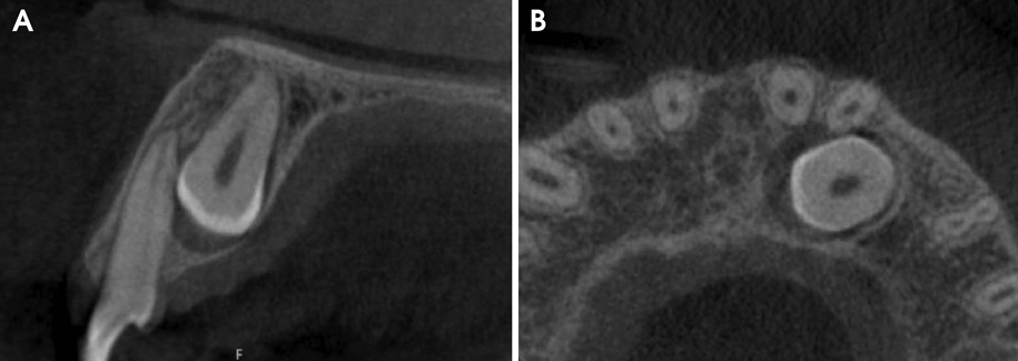
Fig. 1 Sagittal (A) and axial (B) slices of cone-beam computed tomography (CBCT) show an impacted canine causing mild root resorption of the lateral incisor.
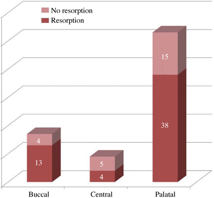
Fig. 2 Distribution of adjacent teeth presenting resorption according to the location of the impacted canines.
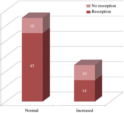
Fig. 3 Distribution of adjacent teeth presenting resorption according to the follicle width of the impacted canines (i.e., normal or increased).
Reference
-
1. Bedoya MM, Park JH. A review of the diagnosis and management of impacted maxillary canines. J Am Dent Assoc. 2009; 140:1485–1493.
Article2. Cooke J, Wang HL. Canine impactions: incidence and management. Int J Periodontics Restorative Dent. 2006; 26:483–491.3. Peck S, Peck L, Kataja M. The palatally displaced canine as a dental anomaly of genetic origin. Angle Orthod. 1994; 64:249–256.4. Baccetti T. Risk indicators and interceptive treatment alternatives for palatally displaced canines. Semin Orthod. 2010; 16:186–192.
Article5. Becker A. In defense of the guidance theory of palatal canine displacement. Angle Orthod. 1995; 65:95–98.6. Walker L, Enciso R, Mah J. Three-dimensional localization of maxillary canines with cone-beam computed tomography. Am J Orthod Dentofacial Orthop. 2005; 128:418–423.
Article7. Sajnani A, King N. Dental age of children and adolescents with impacted maxillary canines. J Orofac Orthop. 2012; 73:359–364.
Article8. Westphalen VP, Gomes de Moraes I, Westphalen FH, Martins WD, Souza PH. Conventional and digital radiographic methods in the detection of simulated external root resorptions: a comparative study. Dentomaxillofac Radiol. 2004; 33:233–235.
Article9. Zhong YL, Zeng XL, Jia QL, Zhang WL, Chen L. Clinical investigation of impacted maxillary canine. Zhonghua Kou Qiang Yi Xue Za Zhi. 2006; 41:483–485.10. Chalakkal P, Thomas AM, Chopra S. Displacement, location, and angulation of unerupted permanent maxillary canines and absence of canine bulge in children. Am J Orthod Dentofacial Orthop. 2011; 139:345–350.
Article11. Mah JK, Alexandroni S. Cone-beam computed tomography in the management of impacted canines. Semin Orthod. 2010; 16:199–204.
Article12. Agrawal JM, Agrawal MS, Nanjannawar LG, Parushetti AD. CBCT in orthodontics: the wave of future. J Contemp Dent Pract. 2013; 14:153–157.13. Scarfe WC, Farman AG, Sukovic P. Clinical applications of cone-beam computed tomography in dental practice. J Can Dent Assoc. 2006; 72:75–80.14. Chen Y, Duan P, Meng Y, Chen Y. Three-dimensional spiral computed tomographic imaging: a new approach to the diagnosis and treatment planning of impacted teeth. Am J Orthod Dentofacial Orthop. 2006; 130:112–116.
Article15. Liu DG, Zhang WL, Zhang ZY, Wu YT, Ma XC. Localization of impacted maxillary canines and observation of adjacent incisor resorption with cone-beam computed tomography. Oral Surg Oral Med Oral Pathol Oral Radiol Endod. 2008; 105:91–98.
Article16. Alqerban A, Jacobs R, Fieuws S, Nackaerts O, Willems G. SEDENTEXCT Project Consortium. Comparison of 6 cone-beam computed tomography systems for image quality and detection of simulated canine impaction-induced external root resorption in maxillary lateral incisors. Am J Orthod Dentofacial Orthop. 2011; 140:e129–e139.
Article17. Bjerklin K, Ericson S. How a computerized tomography examination changed the treatment plans of 80 children with retained and ectopically positioned maxillary canines. Angle Orthod. 2006; 76:43–51.18. Ericson S, Kurol PJ. Resorption of incisors after ectopic eruption of maxillary canines: a CT study. Angle Orthod. 2000; 70:415–423.19. Rozylo-Kalinowska I, Kolasa-Raczka A, Kalinowski P. Dental age in patients with impacted maxillary canines related to the position of the impacted teeth. Eur J Orthod. 2011; 33:492–497.
Article20. Katsnelson A, Flick WG, Susarla S, Tartakovsky JV, Miloro M. Use of panoramic x-ray to determine position of impacted maxillary canines. J Oral Maxillofac Surg. 2010; 68:996–1000.
Article21. Chung DD, Weisberg M, Pagala M. Incidence and effects of genetic factors on canine impaction in an isolated Jewish population. Am J Orthod Dentofacial Orthop. 2011; 139:e331–e335.
Article22. Leuzinger M, Dudic A, Giannopoulou C, Kiliaridis S. Root-contact evaluation by panoramic radiography and cone-beam computed tomography of super-high resolution. Am J Orthod Dentofacial Orthop. 2010; 137:389–392.
Article23. Siegel R, Stós W, Dyras M, Urbanik A, Wojciechowski W, Sztuk S. Assessment of degree and extent of resorption of incisor roots adjacent to impacted maxillary canines. Przegl Lek. 2010; 67:268–274.24. Kim Y, Hyun HK, Jang KT. The position of maxillary canine impactions and the influenced factors to adjacent root resorption in the Korean population. Eur J Orthod. 2011; 34:302–306.
Article25. Postlethwaite KM. Resorption of premolar roots by ectopic canines. Br Dent J. 1989; 167:397–398.
Article26. da Silveira HL, Silveira HE, Liedke GS, Lermen CA, Dos Santos RB, de Figueiredo JA. Diagnostic ability of computed tomography to evaluate external root resorption in vitro. Dentomaxillofac Radiol. 2007; 36:393–396.27. Smailienė D, Sidlauskas A, Lopatienė K, Guzevičienė V, Juodžbalys G. Factors affecting self-eruption of displaced permanent maxillary canines. Medicina (Kaunas). 2011; 47:163–169.
Article28. Ericson S, Bjerklin K, Falahat B. Does the canine dental follicle cause resorption of permanent incisor roots? A computed tomographic study of erupting maxillary canines. Angle Orthod. 2002; 72:95–104.29. Burnett SE. Prevalence of maxillary canine-first premolar transposition in a composite African sample. Angle Orthod. 1999; 69:187–189.30. Ely NJ, Sherriff M, Cobourne MT. Dental transposition as a disorder of genetic origin. Eur J Orthod. 2006; 28:145–151.
Article31. Synodinos PN, Polyzois I. Maxillary canine-first premolar transposition in the permanent dentition: treatment considerations and a case report. J Ir Dent Assoc. 2010; 56:264–267.32. Halazonetis DJ. Horizontally impacted maxillary premolar and bilateral canine transposition. Am J Orthod Dentofacial Orthop. 2009; 135:380–389.
Article33. Liu DG, Zhang WL, Zhang ZY, Wu YT, Ma XC. Three-dimensional evaluations of supernumerary teeth using cone-beam computed tomography for 487 cases. Oral Surg Oral Med Oral Pathol Oral Radiol Endod. 2007; 103:403–411.
Article34. Nagaraj K, Upadhyay M, Yadav S. Impacted maxillary central incisor, canine, and second molar with 2 supernumerary teeth and an odontoma. Am J Orthod Dentofacial Orthop. 2009; 135:390–399.
Article35. European Commission [Internet]. Radiation protection No 172: cone beam CT for dental and maxillofacial radiology (evidencebased guidelines). 2012. cited 2014 May 14. Available from: http://www.sedentexct.eu/files/radiation_protection_172.pdf.36. Rossini G, Cavallini C, Cassetta M, Galluccio G, Barbato E. Localization of impacted maxillary canines using cone beam computed tomography. Ann Stomatol (Roma). 2012; 3:14–18.
- Full Text Links
-
- Actions
-
Cited
- CITED
-
- Close
- Share
-
- Similar articles
-
- Cone beam computed tomography findings of ectopic mandibular third molar in the mandibular condyle: report of a case
- Retrospective Analysis of Incisor Root Resorption Associated with Impacted Maxillary Canines
- Factors Influencing the Duration of Forced Eruption in Impacted Maxillary Canines
- Evaluation of Impacted Maxillary Canine Position Using Panoramic Radiographs and Cone-beam Computed Tomography
- Management of root canal perforation by using cone-beam computed tomography
