Accuracy of linear measurement using cone-beam computed tomography at different reconstruction angles
- Affiliations
-
- 1Department of Dental and Maxillofacial Radiology, School of Dentistry, Shahid Beheshti University of Medical Sciences, Tehran, Iran.
- 2Department of Advanced Periodontology, School of Dentistry, University of Southern California, Los Angeles, USA.
- 3Department of Dental and Maxillofacial Radiology, School of Dentistry, Shahed University of Medical Sciences, Tehran, Iran.
- 4Department of Dental and Maxillofacial Radiology, School of Dentistry, Qazvin University of Medical Sciences, Qazvin, Iran.
- 5Department of Dental and Maxillofacial Radiology, School of Dentistry, Golestan University of Medical Sciences, Golestan, Iran. e_soodeh@yahoo.com
- KMID: 1974493
- DOI: http://doi.org/10.5624/isd.2014.44.4.257
Abstract
- PURPOSE
This study was performed to evaluate the effect of changing the orientation of a reconstructed image on the accuracy of linear measurements using cone-beam computed tomography (CBCT).
MATERIALS AND METHODS
Forty-two titanium pins were inserted in seven dry sheep mandibles. The length of these pins was measured using a digital caliper with readability of 0.01 mm. Mandibles were radiographed using a CBCT device. When the CBCT images were reconstructed, the orientation of slices was adjusted to parallel (i.e., 0degrees), +10degrees, +12degrees, -12degrees, and -10degrees with respect to the occlusal plane. The length of the pins was measured by three radiologists, and the accuracy of these measurements was reported using descriptive statistics and one-way analysis of variance (ANOVA); p<0.05 was considered statistically significant.
RESULTS
The differences in radiographic measurements ranged from -0.64 to +0.06 at the orientation of -12degrees, -0.66 to -0.11 at -10degrees, -0.51 to +0.19 at 0degrees, -0.64 to +0.08 at +10degrees, and -0.64 to +0.1 at +12degrees. The mean absolute values of the errors were greater at negative orientations than at the parallel position or at positive orientations. The observers underestimated most of the variables by 0.5-0.1 mm (83.6%). In the second set of observations, the reproducibility at all orientations was greater than 0.9.
CONCLUSION
Changing the slice orientation in the range of -12degrees to +12degrees reduced the accuracy of linear measurements obtained using CBCT. However, the error value was smaller than 0.5 mm and was, therefore, clinically acceptable.
MeSH Terms
Figure
-
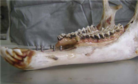
Fig. 1 Titanium pins are inserted in a dried sheep mandible.
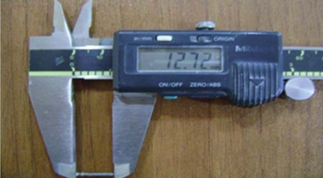
Fig. 2 A digital caliper is used to measure the length of the pins prior to insertion in the mandible.
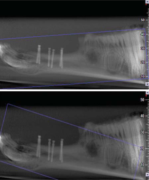
Fig. 3 Scout views show the scans with the reconstruction angle parallel (i.e., at 0°) and at 10° to the occlusal plane.
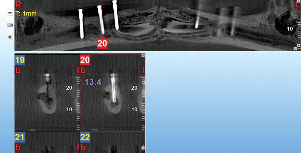
Fig. 4 Linear measurements are performed using the cross-sectional images of the pins.
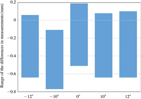
Fig. 5 The graph shows the range of differences in measurements performed by three observers for the different angles used to reconstruct the images.
Cited by 1 articles
-
Intraobserver and interobserver reproducibility in linear measurements on axial images obtained by cone-beam computed tomography
Nathália Cristine da Silva, Maurício Barriviera, José Luiz Cintra Junqueira, Francine Kühl Panzarella, Ricardo Raitz
Imaging Sci Dent. 2017;47(1):11-15. doi: 10.5624/isd.2017.47.1.11.
Reference
-
1. Torres MG, Campos PS, Segundo NP, Navarro M, Crusoé-Rebello I. Accuracy of linear measurements in cone beam computed tomography with different voxel sizes. Implant Dent. 2012; 21:150–155.
Article2. Cavalcanti MG, Rocha SS, Vannier MW. Craniofacial measurements based on 3D-CT volume rendering: implications for clinical applications. Dentomaxillofac Radiol. 2004; 33:170–176.
Article3. Ziegler CM, Woertche R, Brief J, Hassfeld S. Clinical indications for digital volume tomography in oral and maxillofacial surgery. Dentomaxillofac Radiol. 2002; 31:126–130.
Article4. Guerrero ME, Jacobs R, Loubele M, Schutyser F, Suetens P, van Steenberghe D. State-of-the-art on cone beam CT imaging for preoperative planning of implant placement. Clin Oral Investig. 2006; 10:1–7.
Article5. Kau CH, Richmond S, Palomo JM, Hans MG. Three-dimensional cone beam computerized tomography in orthodontics. J Orthod. 2005; 32:282–293.6. Yamamoto K, Ueno K, Seo K, Shinohara D. Development of dento-maxillofacial cone beam X-ray computed tomography system. Orthod Craniofac Res. 2003; 6:Suppl 1. 160–162.
Article7. Scarfe WC, Farman AG, Sukovic P. Clinical applications of cone-beam computed tomography in dental practice. J Can Dent Assoc. 2006; 72:75–80.8. Kumar V, Ludlow JB, Mol A, Cevidanes L. Comparison of conventional and cone beam CT synthesized cephalograms. Dentomaxillofac Radiol. 2007; 36:263–269.
Article9. Li T, MacDonald D. Osseo integrated implants. In : MacDonald D, editor. Oral and maxillofacial radiology: a diagnostic approach. Chichester: Wiley-Blackwell;2011. p. 296–298.10. Boas FE, Fleischmann D. CT artifact: cause and reduction techniques. Imaging Med. 2012; 4:229–240.11. Ganguly R, Ruprecht A, Vincent S, Hellstein J, Timmons S, Qian F. Accuracy of linear measurement in the Galileos cone beam computed tomography under simulated clinical conditions. Dentomaxillofac Radiol. 2011; 40:299–305.
Article12. Sheikhi M, Ghorbanizadeh S, Abdinian M, Goroohi H, Badrian H. Accuracy of linear measurements of Galileos cone beam computed tomography in normal and different head positions. Int J Dent. 2012; 2012:214954.
Article13. Tomasi C, Bressan E, Corazza B, Mazzoleni S, Stellini E, Lith A. Reliability and reproducibility of linear mandible measurements with the use of a cone-beam computed tomography and two object inclinations. Dentomaxillofac Radiol. 2011; 40:244–250.
Article14. Bashizadeh Fakhar H, Abbaszadeh A. Effect of mandibular plane angle on image dimensions in linear tomography. J Dent Med. 2011; 24:42–49.15. Hassan B, van der Stelt P, Sanderink G. Accuracy of three-dimensional measurements obtained from cone beam computed tomography surface-rendered images for cephalometric analysis: influence of patient scanning position. Eur J Orthod. 2009; 31:129–134.
Article16. Moshiri M, Scarfe WC, Hilgers ML, Scheetz JP, Silveira AM, Farman AG. Accuracy of linear measurements from imaging plate and lateral cephalometric images derived from cone-beam computed tomography. Am J Orthod Dentofacial Orthop. 2004; 132:550–560.
Article17. Ludlow JB, Laster WS, See M, Bailey LJ, Hershey HG. Accuracy of measurements of mandibular anatomy in cone beam computed tomography images. Oral Surg Oral Med Oral Pathol Oral Radiol Endod. 2007; 103:534–542.
Article18. Moshfeghi M, Tavakoli MA, Hosseini ET, Hosseini AT, Hosseini IT. Analysis of linear measurement accuracy obtained by cone beam computed tomography (CBCT-NewTom VG). Dent Res J (Isfahan). 2012; 9:S57–S62.
- Full Text Links
-
- Actions
-
Cited
- CITED
-
- Close
- Share
-
- Similar articles
-
- Comparison of cone-beam computed tomography cephalometric measurements using a midsagittal projection and conventional two-dimensional cephalometric measurements
- Management of root canal perforation by using cone-beam computed tomography
- The accuracy of the imaging reformation of cone beam computed tomography for the assessment of bone defect healing
- Evaluation of linear measurements of implant sites based on head orientation during acquisition: An ex vivo study using cone-beam computed tomography
- Linear accuracy of cone-beam computed tomography and a 3-dimensional facial scanning system: An anthropomorphic phantom study
