Imaging Sci Dent.
2014 Sep;44(3):221-227. 10.5624/isd.2014.44.3.221.
Cone-beam computed tomography analysis of root and canal morphology of mandibular premolars in a Spanish population
- Affiliations
-
- 1Department of Stomatology, Universitat de Valencia, Valencia, Spain. llena@uv.es
- 2Fernandez Ugedo y Chaves Clinic, Alicante, Spain.
- KMID: 1974486
- DOI: http://doi.org/10.5624/isd.2014.44.3.221
Abstract
- PURPOSE
This study aimed to investigate the clinical anatomy of lower premolar roots in a Spanish population by using cone-beam computed tomography (CBCT), correlating findings with patient gender and tooth type.
MATERIALS AND METHODS
Using 70 CBCT images, we evaluated 126 healthy, untreated, well-developed lower premolars. The number and morphology of roots and root canals, and the foramina number were assessed. Results for gender and tooth type were compared using the chi-squared and ANOVA tests.
RESULTS
The average length of teeth and roots was significantly higher in men (p=0.00). All 126 premolars had a single root. One canal was found in 83.3% of the premolars, with no gender or tooth type differences; Vertucci configuration types I and V were the most prevalent. The first premolars showed significantly greater variability than the second premolars (p=0.03). A single apical foramen was found in 89.7% of the premolars, with no differences by tooth type. Women had a significantly higher prevalence of two apical foramina than men (p=0.04). Some degree of curvature was observed in 65% of the premolars, with no differences by gender or tooth type. A root angle of more than 20degrees was found in 12.98% of the premolars, without any differences by gender or tooth.
CONCLUSION
All premolars were single-rooted. One canal had the most prevalent morphology. More variability in canal anatomy was found in the first premolars. Curvatures greater than 20degrees were found at less than 5 mm from the apex.
MeSH Terms
Figure
-
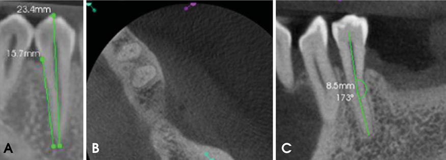
Fig. 1 A. Total tooth length from the cuspid to the apex and root length from the cemento-enamel junction to the apex are measured using a mesiodistal cone-beam computed tomography (CBCT) image. B. Axial CBCT image shows two premolars with two canals. C. Root curvature and distance from the angle vertex to the apex are measured using a mesiodistal CBCT image.
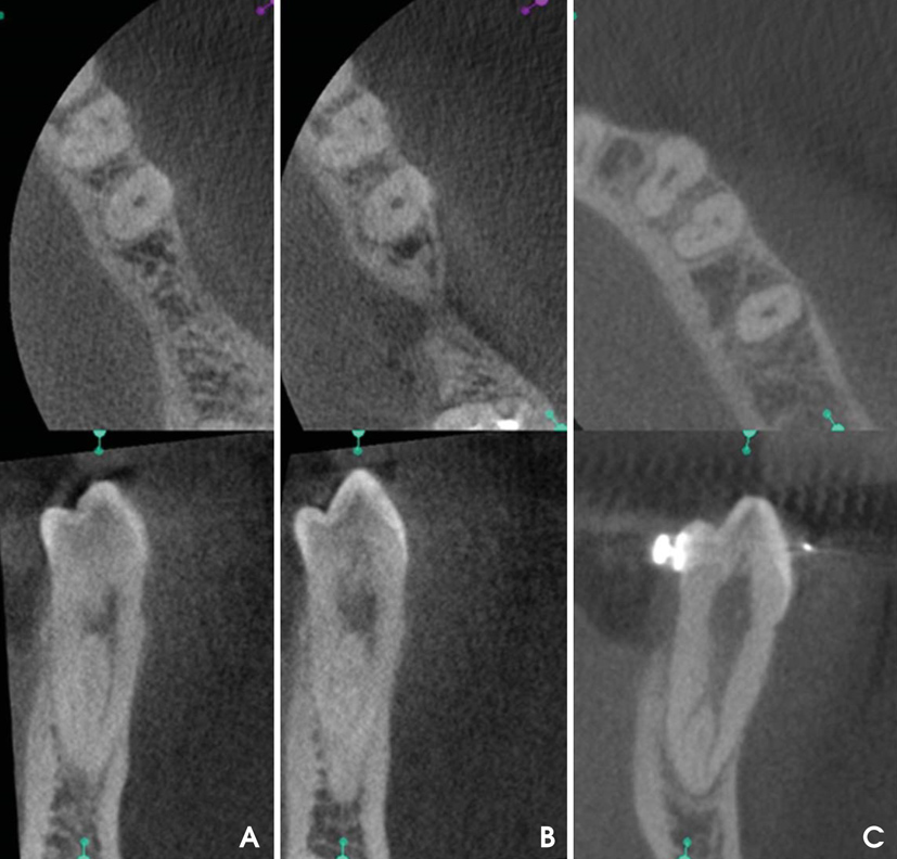
Fig. 2 Axial (up) and buccolingual (down) CBCT images show the Vertucci type II configuration. B. Shapes not included in Vertucci's classification. One canal in the coronal third of the root, three canals in the middle third, and one canal in the apical third. C. Axial (up) and buccolingual (down) CBCT images show the Vertucci type V configuration.
Reference
-
1. Miracle AC, Mukherji SK. Conebeam CT of the head and neck, part 2: clinical applications. AJNR Am J Neuroradiol. 2009; 30:1285–1292.
Article2. Yajima A, Otonari-Yamamoto M, Sano T, Hayakawa Y, Otonari T, Tanabe K, et al. Cone-beam CT (CB Throne) applied to dentomaxillofacial region. Bull Tokyo Dent Coll. 2006; 47:133–141.
Article3. Scarfe WC, Farman AG, Sukovic P. Clinical applications of cone-beam computed tomography in dental practice. J Can Dent Assoc. 2006; 72:75–80.4. Madrigal C, Ortega R, Meniz C, López-Quiles J. Study of available bone for interforaminal implant treatment using conebeam computed tomography. Med Oral Patol Oral Cir Bucal. 2008; 13:E307–E312.5. Scarfe WC, Levin MD, Gane D, Farman AG. Use of cone beam computed tomography in endodontics. Int J Dent. 2009; 2009:634567.
Article6. Patel S. New dimensions in endodontic imaging: part 2. Cone beam computed tomography. Int Endod J. 2009; 42:463–475.
Article7. Patel S, Horner K. The use of cone beam computed tomography in endodontics. Int Endod J. 2009; 42:755–756.
Article8. Zillich R, Dowson J. Root canal morphology of mandibular first and second premolars. Oral Surg Oral Med Oral Pathol. 1973; 36:738–744.
Article9. Slowey RR. Root canal anatomy. Road map to successful endodontics. Dent Clin North Am. 1979; 23:555–573.10. Glassman GD. Flare-up with associated paresthesia of a mandibular second premolar with three root canals. Oral Surg Oral Med Oral Pathol. 1987; 64:110–113.
Article11. Cleghorn BM, Christie WH, Dong CC. The root and root canal morphology of the human mandibular first premolar: a literature review. J Endod. 2007; 33:509–516.
Article12. Cleghorn BM, Christie WH, Dong CC. The root and root canal morphology of the human mandibular second premolar: a literature review. J Endod. 2007; 33:1031–1037.
Article13. Rahimi S1, Shahi S, Yavari HR, Manafi H, Eskandarzadeh N. Root canal configuration of mandibular first and second premolars in an Iranian population. J Dent Res Dent Clin Dent Prospects. 2007; 1:59–64.14. Park JB, Kim N, Park S, Kim Y, Ko Y. Evaluation of root anatomy of permanent mandibular premolars and molars in a Korean population with cone-beam computed tomography. Eur J Dent. 2013; 7:94–101.15. Trope M, Elfenbein L, Tronstad L. Mandibular premolars with more than one root canal in different race groups. J Endod. 1986; 12:343–345.
Article16. Hulsmann M. Mandibular first premolar with three root canals. Endod Dent Traumatol. 1990; 6:189–191.
Article17. Yang ZP. Multiple canals in a mandibular first premolar. Case report. Aust Dent J. 1994; 39:18–19.
Article18. Nallapati S. Three canal mandibular first and second premolars: a treatment approach. J Endod. 2005; 31:474–476.19. Kakkar P, Singh A. Mandibular first premolar with three roots: a case report. Iran Endod J. 2012; 7:207–210.20. Yu X, Guo B, Li KZ, Zhang R, Tian YY, Wang H, et al. Conebeam computed tomography study of root and canal morphology of mandibular premolars in a western Chinese population. BMC Med Imaging. 2012; 12:18.
Article21. Michetti J, Maret D, Mallet JP, Diemer F. Validation of cone beam computed tomography as a tool to explore root canal anatomy. J Endod. 2010; 36:1187–1190.
Article22. Vertucci FJ. Root canal anatomy of the human permanent teeth. Oral Surg Oral Med Oral Pathol. 1984; 58:589–599.
Article23. Khedmat S, Assadian H, Saravani AA. Root canal morphology of the mandibular first premolars in an Iranian population using cross-sections and radiography. J Endod. 2010; 36:214–217.
Article24. Tzanetakis GN, Lagoudakos TA, Kontakiotis EG. Endodontic treatment of a mandibular second premolar with four canals using operating microscope. J Endod. 2007; 33:318–321.
Article25. Awawdeh LA, Al-Qudah AA. Root form and canal morphology of mandibular premolars in a Jordanian population. Int Endod J. 2008; 41:240–248.
Article26. Kim Y, Lee SJ, Woo J. Morphology of maxillary first and second molars analyzed by cone-beam computed tomography in a Korean population: variations in the number of roots and canals and the incidence of fusion. J Endod. 2012; 38:1063–1068.
Article27. Zhang R, Yang H, Yu X, Wang H, Hu T, Dummer PM. Use of CBCT to identify the morphology of maxillary permanent molar teeth in a Chinese subpopulation. Int Endod J. 2011; 44:162–169.
Article28. Zheng Q, Zhang L, Zhou X, Wang Q, Wang Y, Tang L, et al. C-shaped root canal system in mandibular second molars in a Chinese population evaluated by cone-beam computed tomography. Int Endod J. 2011; 44:857–862.
Article29. Reis AG, Grazziotin-Soares R, Barletta FB, Fontanella VR, Mahl CR. Second canal in mesiobuccal root of maxillary molars is correlated with root third and patient age: a cone-beam computed tomographic study. J Endod. 2013; 39:588–592.
Article30. Demirbuga S, Sekerci AE, Dinçer AN, Cayabatmaz M, Zorba YO. Use of cone-beam computed tomography to evaluate root and canal morphology of mandibular first and second molars in Turkish individuals. Med Oral Patol Oral Cir Bucal. 2013; 18:e737–e744.
Article31. Silva EJ, Nejaim Y, Silva AV, Haiter-Neto F, Cohenca N. Evaluation of root canal configuration of mandibular molars in a Brazilian population by using cone-beam computed tomography: an in vivo study. J Endod. 2013; 39:849–852.32. Kim E, Fallahrastegar A, Hur YY, Jung IY, Kim S, Lee SJ. Difference in root canal length between Asians and Caucasians. Int Endod J. 2005; 38:149–151.
Article33. Vertucci FJ. Root canal morphology of mandibular premolars. J Am Dent Assoc. 1978; 97:47–50.
Article34. Green D. Double canals in single roots. Oral Surg Oral Med Oral Pathol. 1973; 35:689–696.
Article35. Lu TY, Yang SF, Pai SF. Complicated root canal morphology of mandibular first premolar in a Chinese population using the cross section method. J Endod. 2006; 32:932–936.
Article36. Willershausen B, Kasaj A, Röhrig B, Briseño B. The determination of the initial straight length in root canals of mandibular premolars - an in vitro study. Eur J Med Res. 2009; 14:85–89.37. Lombart B, Michonneau JC. Premolar anatomy and endodontic treatment. Rev Belge Med Dent (1984). 2005; 60:322–336.
- Full Text Links
-
- Actions
-
Cited
- CITED
-
- Close
- Share
-
- Similar articles
-
- Observation of mandibular second molar roots and root canal morphology using dental cone-beam computed tomography
- Characterization of mandibular molar root and canal morphology using cone beam computed tomography and its variability in Belgian and Chilean population samples
- Prevalence and features of distolingual roots in mandibular molars analyzed by cone-beam computed tomography
- Cone-beam computed tomography observation of maxillary first premolar canal shapes
- Reliability of panoramic radiography in predicting proximity of third molars to the mandibular canal: A comparison using cone-beam computed tomography
