Cone-beam computed tomography observation of maxillary first premolar canal shapes
- Affiliations
-
- 1Department of Morphological Biology, Ohu University School of Dentistry, Koriyama, Japan
- 2Department of General Odontology, Ohu University Graduate School of Dentistry, Koriyama, Japan
- 3Department of Oral Radiology and Diagnosis, Ohu University School of Dentistry, Koriyama, Japan
- KMID: 2523572
- DOI: http://doi.org/10.5115/acb.21.110
Abstract
- A variety of shapes has been reported for the roots and root canals of maxillary first premolars. The purposes of the present study were to determine branching and shapes of the roots of maxillary first premolars, as well as age-related changes using slice images obtained with cone-beam computed tomography (CBCT) for dental use. CBCT-reconstructed images of 125 cases that included maxillary first premolars were used as subjects. Slice images at the cervical one-third, center, and apical one-third positions of the root were prepared. Root branching and number of root canals was determined at each measurement position in the images. The subjects were divided into three groups: younger than 30 years, 30 to 50 years, and over 50 years. The root canal morphology was compared among these age groups. Single-rooted premolars were the most frequent. As for number of root canals, a single-canal premolar was observed at the position of the cervical one-third in 33.6%, at the center in 35.2%, and at the apical one-third in 56.0%. Thereafter the subjects were divided into groups by age, namely, younger than 30 years, 30 to 50 years, and over 50 years old, and it was revealed that the ratio of the two-canal type increased with age. In regard to tooth morphology, it was confirmed that the two-canal type shows more frequent occurrence with aging in maxillary first premolar. Based on our findings, we consider that CBCT can be useful for determining the root canal morphology with complicated shapes.
Figure
-

Fig. 1 The axial slice images of cone-beam computed tomography (CBCT) revealed different numbers of roots of the maxillary first premolars. Slice images at cervical one-third (A–C), root center (D–F), and apical one-third (G–I). Slice images of one root (A, D, G), two roots (B, E, H), and three roots (C, F, I).
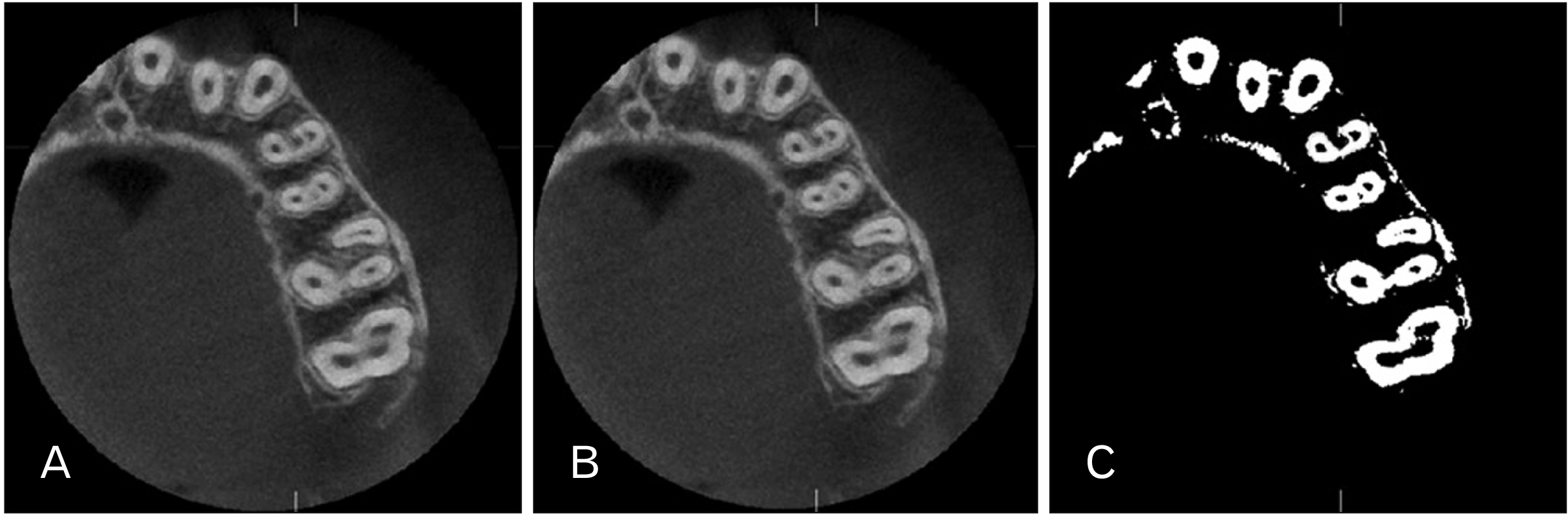
Fig. 2 Binary image processing. (A) Slice image of root center of left maxillary first premolar. (B) Image analyzed using peri-root portion as area of interest. (C) Binary image of area of interest.
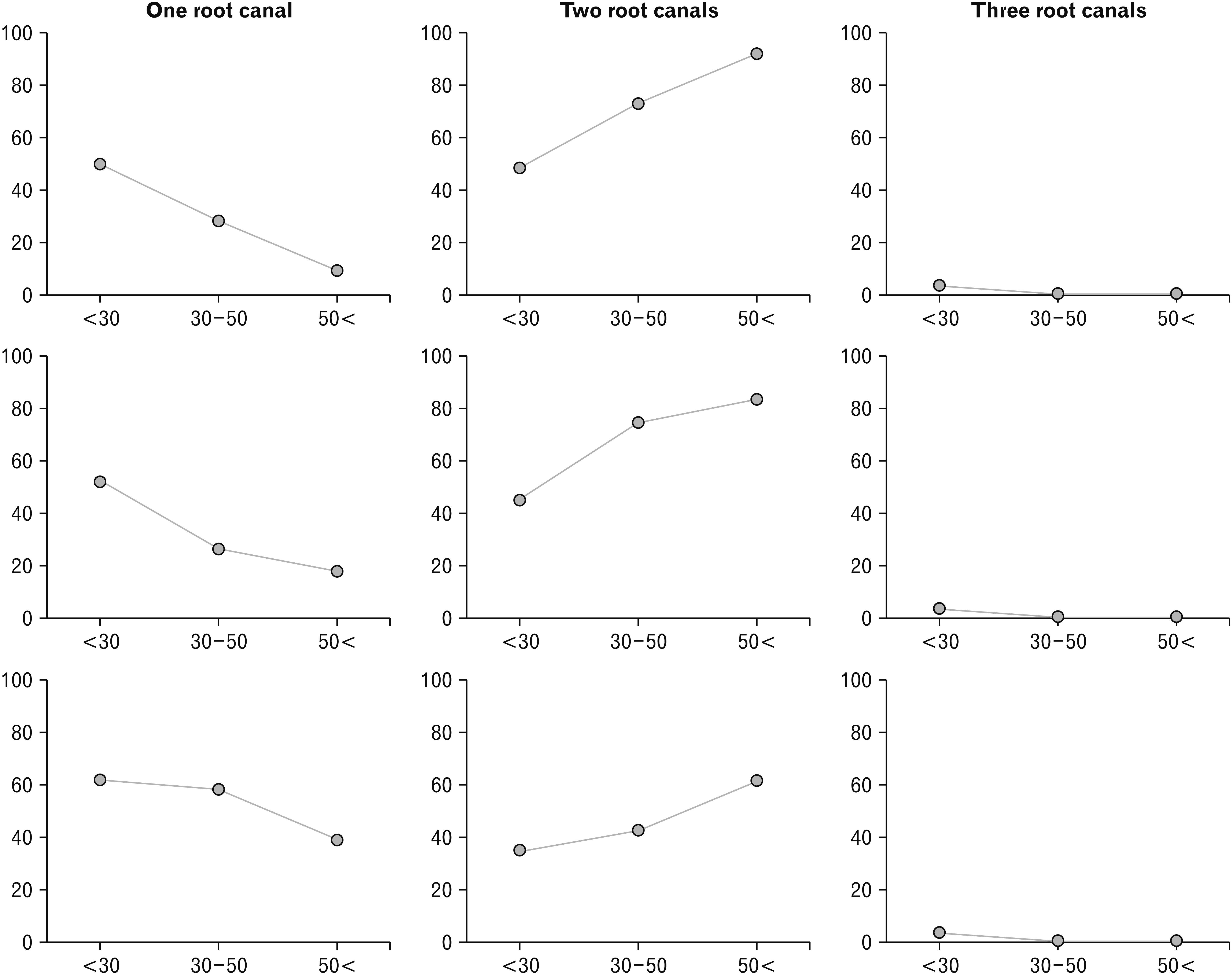
Fig. 3 Root canal numbers of the maxillary first premolars divided by age. Left: Occurrence of single canal. Center: Occurrence of two canals. Right: Occurrence of three canals. The shape at the cervical one-third, root center, and apical one-third is shown in the upper, middle, and bottom, respectively. Charts show occurrence after dividing into age groups (from the left: <30, 30–50, 50< years old).
Cited by 3 articles
-
Observation of mandibular second molar roots and root canal morphology using dental cone-beam computed tomography
Ryohei Shigefuji, Masamitsu Serikawa, Akinobu Usami
Anat Cell Biol. 2022;55(2):155-160. doi: 10.5115/acb.22.050.Accuracy verification of dental cone-beam computed tomography of mandibular incisor root canals and assessment of its morphology and aging-related changes
Katsuyuki Aoki, Masamitsu Serikawa, Takuya Harada, Akinobu Usami
Anat Cell Biol. 2023;56(2):185-190. doi: 10.5115/acb.22.247.Dental characteristics on panoramic radiographs as parameters for non-invasive age estimation: a pilot study
Harin Cheong, Akiko Kumagai, Sehyun Oh, Sang-Seob Lee
Anat Cell Biol. 2023;56(4):474-481. doi: 10.5115/acb.23.140.
Reference
-
References
1. Vertucci FJ, Gegauff A. 1979; Root canal morphology of the maxillary first premolar. J Am Dent Assoc. 99:194–8. DOI: 10.14219/jada.archive.1979.0255. PMID: 287737.
Article2. Vertucci FJ. 1984; Root canal anatomy of the human permanent teeth. Oral Surg Oral Med Oral Pathol. 58:589–99. DOI: 10.1016/0030-4220(84)90085-9. PMID: 6595621.
Article3. Neelakantan P, Subbarao C, Ahuja R, Subbarao CV. 2011; Root and canal morphology of Indian maxillary premolars by a modified root canal staining technique. Odontology. 99:18–21. DOI: 10.1007/s10266-010-0137-0. PMID: 21271321.
Article4. Lipski M, Wozniak K, Lagocka R, Tomasik M. 2005; Root and canal morphology of the first human maxillary premolar. Durham Anthropol J. 12:2–3.5. Pineda F, Kuttler Y. 1972; Mesiodistal and buccolingual roentgenographic investigation of 7,275 root canals. Oral Surg Oral Med Oral Pathol. 33:101–10. DOI: 10.1016/0030-4220(72)90214-9. PMID: 4500261.
Article6. Soares JA, Leonardo RT. 2003; Root canal treatment of three-rooted maxillary first and second premolars--a case report. Int Endod J. 36:705–10. DOI: 10.1046/j.1365-2591.2003.00711.x. PMID: 14596244.7. Sieraski SM, Taylor GN, Kohn RA. 1989; Identification and endodontic management of three-canalled maxillary premolars. J Endod. 15:29–32. DOI: 10.1016/S0099-2399(89)80095-0. PMID: 2607264.
Article8. Javidi M, Zarei M, Vatanpour M. 2008; Endodontic treatment of a radiculous maxillary premolar: a case report. J Oral Sci. 50:99–102. DOI: 10.2334/josnusd.50.99. PMID: 18403892.
Article9. Slowey RR. 1979; Root canal anatomy. Road map to successful endodontics. Dent Clin North Am. 23:555–73. PMID: 294389.10. Beltes P, Kalaitzoglou ME, Kantilieraki E, Beltes C, Angelopoulos C. 2017; 3-rooted maxillary first premolars: an ex vivo study of external and internal morphologies. J Endod. 43:1267–72. DOI: 10.1016/j.joen.2017.03.045. PMID: 28662880.11. Abella F, Teixidó LM, Patel S, Sosa F, Duran-Sindreu F, Roig M. 2015; Cone-beam computed tomography analysis of the root canal morphology of maxillary first and second premolars in a Spanish population. J Endod. 41:1241–7. DOI: 10.1016/j.joen.2015.03.026. PMID: 25956606.12. Berman LH, Hartwell GR. Hargreaves KM, Cohen S, editors. 2011. Diagnosis. Cohen's Pathways of the Pulp. 10th ed. Elsevier;St. Louis: p. 2–39. DOI: 10.1016/B978-0-323-06489-7.00001-1. PMID: 21303513. PMCID: PMC3045897.13. Suomalainen A, Pakbaznejad Esmaeili E, Robinson S. 2015; Dentomaxillofacial imaging with panoramic views and cone beam CT. Insights Imaging. 6:1–16. DOI: 10.1007/s13244-014-0379-4. PMID: 25575868. PMCID: PMC4330237.
Article14. Schneider CA, Rasband WS, Eliceiri KW. 2012; NIH image to ImageJ: 25 years of image analysis. Nat Methods. 9:671–5. DOI: 10.1038/nmeth.2089. PMID: 22930834. PMCID: PMC5554542.
Article15. Special Committee to Revise the Joint AAE/AAOMR Position Statement on use of CBCT in Endodontics. 2015; AAE and AAOMR joint position statement: use of cone beam computed tomography in endodontics 2015 update. Oral Surg Oral Med Oral Pathol Oral Radiol. 120:508–12. DOI: 10.1016/j.oooo.2015.07.033. PMID: 26346911.16. Katakami K, Mishima A, Shiozaki K, Shimoda S, Hamada Y, Kobayashi K. 2008; Characteristics of accessory mental foramina observed on limited cone-beam computed tomography images. J Endod. 34:1441–5. DOI: 10.1016/j.joen.2008.08.033. PMID: 19026870.
Article17. Kim SY, Yang SE. 2012; Cone-beam computed tomography study of incidence of distolingual root and distance from distolingual canal to buccal cortical bone of mandibular first molars in a Korean population. J Endod. 38:301–4. DOI: 10.1016/j.joen.2011.10.023. PMID: 22341064.
Article18. Blattner TC, George N, Lee CC, Kumar V, Yelton CD. 2010; Efficacy of cone-beam computed tomography as a modality to accurately identify the presence of second mesiobuccal canals in maxillary first and second molars: a pilot study. J Endod. 36:867–70. DOI: 10.1016/j.joen.2009.12.023. PMID: 20416435.
Article19. Vizzotto MB, Silveira PF, Arús NA, Montagner F, Gomes BP, da Silveira HE. 2013; CBCT for the assessment of second mesiobuccal (MB2) canals in maxillary molar teeth: effect of voxel size and presence of root filling. Int Endod J. 46:870–6. DOI: 10.1111/iej.12075. PMID: 23442087.
Article20. Morikage N, Hamada T, Usami A, Takada S. 2017; Topographical relationship between positions of lingual foramina and attachment of mylohyoid muscle in mental region. Surg Radiol Anat. 39:735–9. DOI: 10.1007/s00276-016-1804-9. PMID: 28078367.
Article21. Bulut DG, Kose E, Ozcan G, Sekerci AE, Canger EM, Sisman Y. 2015; Evaluation of root morphology and root canal configuration of premolars in the Turkish individuals using cone beam computed tomography. Eur J Dent. 9:551–7. DOI: 10.4103/1305-7456.172624. PMID: 26929695. PMCID: PMC4745238.
Article22. George R, Rutley EB, Walsh LJ. 2008; Evaluation of smear layer: a comparison of automated image analysis versus expert observers. J Endod. 34:999–1002. DOI: 10.1016/j.joen.2008.05.003. PMID: 18634934.
Article23. Schmidt TF, Teixeira CS, Felippe MC, Felippe WT, Pashley DH, Bortoluzzi EA. 2015; Effect of ultrasonic activation of irrigants on smear layer removal. J Endod. 41:1359–63. DOI: 10.1016/j.joen.2015.03.023. PMID: 25960002.24. Nelson S. Nelson S, editor. 2015. Pulp chambers and canals. Wheeler's Dental Anatomy, Physiology and Occlusion. 10th ed. Elsevier;St. Louis: p. 203–30.25. Tian YY, Guo B, Zhang R, Yu X, Wang H, Hu T, Dummer PM. 2012; Root and canal morphology of maxillary first premolars in a Chinese subpopulation evaluated using cone-beam computed tomography. Int Endod J. 45:996–1003. DOI: 10.1111/j.1365-2591.2012.02059.x. PMID: 22551454.
Article26. Berkovitz BKB. Standring S, editor. 2016. Oral cavity. Gray's Anatomy. 41st ed. Elsevier;Livingstone: p. 507–33.
Article27. Nelson S. Nelson S, editor. 2015. Development and eruption of the teeth. Wheeler's Dental Anatomy, Physiology and Occlusion. 10th ed. Elsevier;St. Louis: p. 21–42.28. Fan L, Yuan K, Niu C, Ma R, Huang Z. 2018; A cone-beam computed tomography study of the mesial cervical concavity of maxillary first premolars. Arch Oral Biol. 92:79–82. DOI: 10.1016/j.archoralbio.2018.05.002. PMID: 29775860.
Article29. Sicher H, Dubrul EL. Sicher H, DuBrul EL, editors. 1975. The viscera. Oral Anatomy. 6th ed. Mosby;St Louis: p. 192–298.
- Full Text Links
-
- Actions
-
Cited
- CITED
-
- Close
- Share
-
- Similar articles
-
- Detection of maxillary second molar with two palatal roots using cone beam computed tomography: a case report
- A cone-beam computed tomographic study of C-shaped root and root canal in maxillary molars
- Distribution of Vascular Canal at Lateral Wall of Maxillary Sinus in Korean Population Using Computed Tomography
- Management of root canal perforation by using cone-beam computed tomography
- Analysis of the root position and angulation of maxillary premolars in alveolar bone using cone-beam computed tomography
