Ultrasonographic findings of mesenchymal chondrosarcoma of the mandible: report of a case
- Affiliations
-
- 1Biomaterial Research center, Oral Radiology Department, Dental School, Shiraz University of Medical Sciences, Shiraz, Iran.
- 2TABA Medical Imaging Center, Shiraz, Iran.
- 3Oral Surgery Department, Dental School, Shiraz University of Medical Sciences, Shiraz, Iran.
- 4Oral Radiology Department, Dental School, Shiraz University of Medical Sciences, Shiraz, Iran. maneli.houshyar@yahoo.com
- 5Dental Center of Dastgheib Hospital, Shiraz, Iran.
- 6Oral Pathology Department, Dental School, Shiraz University of Medical Sciences, Shiraz, Iran.
- KMID: 1974419
- DOI: http://doi.org/10.5624/isd.2012.42.2.115
Abstract
- Today, ultrasound imaging is being widely used to assess soft tissue lesions in the maxillofacial region. However, ultrasound investigations of intra-osseous lesions are rare, especially for tumors of the jaws. This report emphasized the capability of this useful imaging modality in identification of the characteristics of malignant conditions involving the bone. Mesenchymal chondrosarcoama, one of the unusual malignant conditions of the jaw, was presented in a young male with significant facial swelling. Different imaging modalities parallel with the histopathologic investigation confirmed the diagnosis. Interestingly, destruction of the bony cortex and new bone formation with a characteristic "sun ray appearance", highly suggestive of sarcomas, was manifested on the ultrasonograph. Thus, this report presented the ultrasonographic features of chondrosarcoma of mandible and considered the ultrasonography to be a useful imaging modality to evaluate intra-osseous jaw lesions.
MeSH Terms
Figure
-

Fig. 1 Facial photographs reveal a large facial swelling on left side.
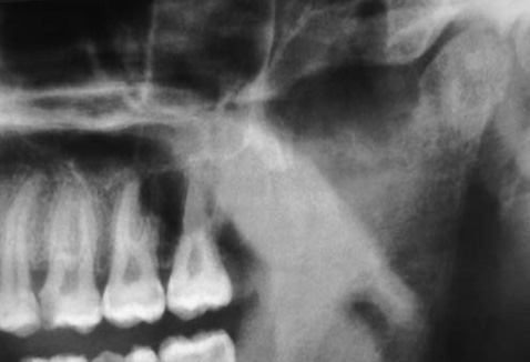
Fig. 2 Cropped panoramic radiograph reveals destruction of cortical bone in the region of left sigmoid notch and ascending ramus. Irregularity of cortical border in the region is observed.
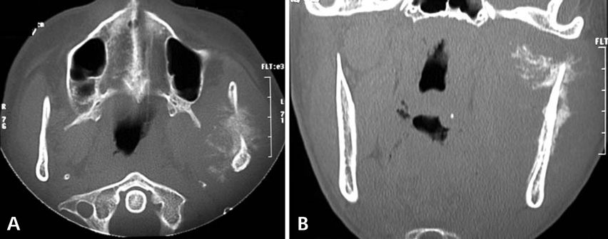
Fig. 3 Axial CT (A) and coronal CT (B) images show a destructive lesion with spiculated periosteal new bone formation. New bone spicules are perpendicular to the original cortex, giving the so-called "sun ray" or "sun burst" appearance.

Fig. 4 Ultrasonographic image shows soft tissue component of the lesion with well-defined hypo-echogenic pattern and hard tissue component of the lesion with ill-defined hyper-echoic pattern. B, C, and D. Ultrasonographic images reveal the characteristic "sun burst" appearance of new bone spicules perpendicular to outer cortical plate which is consistent with CT findings.

Fig. 5 A. Histopathologic examination shows tumor mass composed of sheets of dysplastic spindle cells within a chondroid matrix (H&E stain, ×100). B. Malignant chondroid matrix with areas of calcification is manifested (H&E stain, ×400).
Reference
-
1. Pontes HA, Pontes FS, de Abreu MC, de Carvalho PL, de Brito Kato AM, Fonseca FP, et al. Clinicopathological analysis of head and neck chondrosarcoma: three case reports and literature review. Int J Oral Maxillofac Surg. 2012. 41:203–210.
Article2. White SC, Pharoah MJ. Oral radiology; principles and interpretation. 2009. 6th ed. St. Louis: Mosby Elsevier.3. Ruark DS, Schlehaider UK, Shah JP. Chondrosarcomas of the head and neck. World J Surg. 1992. 16:1010–1016.
Article4. Sammartino G, Marenzi G, Howard CM, Minimo C, Trosino O, Califano L, et al. Chondrosarcoma of the jaw: a closer look at its management. J Oral Maxillofac Surg. 2008. 66:2349–2355.
Article5. Ariyoshi Y, Shimahara M. Mesenchymal chondrosarcoma of the maxilla: report of a case. J Oral Maxillofac Surg. 1999. 57:733–737.
Article6. Sharvit A, Gutman D, Laufer D, Robinson E. Correlation between bone scanning and the radiographic image in the diagnosis of osteosarcoma. Int J Oral Surg. 1975. 4:172–176.
Article7. Myers RP, Jiang S, Girotto JA. A rare tumour in the mandible of a young man: a case report and literature review of mesenchymal chondrosarcoma of the mandible. Oral Surg. 2009. 2:29–35.
Article8. Zakkak TB, Flynn TR, Boguslaw B, Adamo AK. Mesenchymal chondrosarcoma of the mandible: case report and review of the literature. J Oral Maxillofac Surg. 1998. 56:84–91.
Article9. Ng SY, Songra A, Ali N, Carter JL. Ultrasound features of osteosarcoma of the mandible - a first report. Oral Surg Oral Med Oral Pathol Oral Radiol Endod. 2001. 92:582–586.10. Lu L, Yang J, Liu JB, Yu Q, Xu Q. Ultrasonographic evaluation of mandibular ameloblastoma: a preliminary observation. Oral Surg Oral Med Oral Pathol Oral Radiol Endod. 2009. 108:e32–e38.
Article
- Full Text Links
-
- Actions
-
Cited
- CITED
-
- Close
- Share
-
- Similar articles
-
- Mesenchymal chondrosarcoma on right mandible: a case report
- Mesenchymal Chondrosarcoma of the Orbit: A case report and review of the literature
- Mesenchymal Chondrosarcoma Involving Posterior Bone of C7, T1, T2 and Adjacent Muscles
- Mesenchymal Chondrosarcoma: A Case Report
- Fine Needle Aspiration Cytology of Extraskeletal Mesenchymal Chondrosarcoma
