Imaging Sci Dent.
2012 Jun;42(2):77-81. 10.5624/isd.2012.42.2.77.
Odontoma: a retrospective study of 73 cases
- Affiliations
-
- 1Department of Oral and Maxillofacial Radiology, School of Dentistry, Kyungpook National University, Daegu, Korea. chan@knu.ac.kr
- KMID: 1974412
- DOI: http://doi.org/10.5624/isd.2012.42.2.77
Abstract
- PURPOSE
The purpose of the present study was to retrospectively evaluate the clinical findings and treatment results for impacted permanent teeth associated with odontomas.
MATERIALS AND METHODS
We retrospectively investigated 73 odontomas in 72 patients who visited Kyungpook National University Dental Hospital from April 2004 through November 2011. The study was performed using medical records, panoramic radiographs, and pathological reports. Data gathered included age, gender, location, chief complaints, effects on dentition, and treatment of odontoma and the impacted tooth associated with odontoma.
RESULTS
Most compound odontomas (46.7%) were found in the second decade and complex odontomas were not related to age. Odontomas showed no gender predilection. Fifty-five cases (75.3%) of odontomas were detected on routine dental radiographs. Sixty percent of compound odontomas occurred in the canine area and 57.1% of complex odontomas in the molar areas. Impaction of permanent teeth (61.6%) was the most common complication on the adjacent teeth. Most odontomas (84.9%) were removed surgically and impacted permanent teeth were managed by surgical removal (53.2%), orthodontic treatment (25.5%), or surgical repositioning (6.4%). There was a statistically significant relation between age and preservation of the impacted permanent teeth associated with odontomas (p<0.01).
CONCLUSION
Early detection and treatment of odontomas increase the possibility of preservation of the impacted tooth. Therefore, it would be suggested that periodic panoramic examination during the first and second decade of life might be beneficial for the early detection and better prognosis of odontomas.
Keyword
MeSH Terms
Figure
-
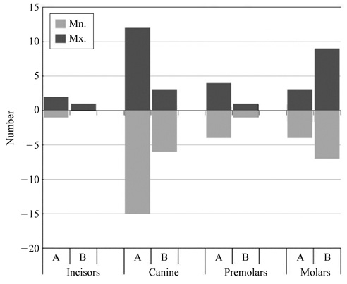
Fig. 1 Location of odontomas (A; compound type, B; complex type).
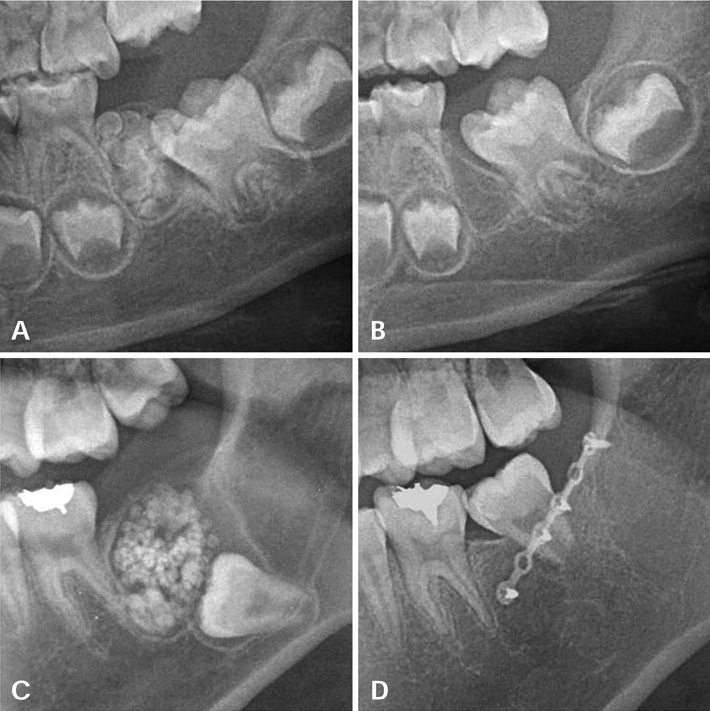
Fig. 2 Pre- (A) and post-operative (3 months follow-up) (B) cropped panoramic radiographs reveal the compound odontoma and normal eruption of impacted tooth after excision of odontoma mass. Cropped panoramic radiographs show preoperative compound odontoma (C) and surgically repositioned impacted tooth after excision (7 months follow-up) (D). (A. 5 years 6 months, B. 6 years, C. 17 years 6 months, D. 18 years 1 month).
Cited by 1 articles
-
Giant complex odontoma in the posterior mandible: A case report and literature review
Jong Chan Park, Ji Ho Yang, Sung Youn Jo, Bong Chul Kim, Jun Lee, Wan Lee
Imaging Sci Dent. 2018;48(4):289-293. doi: 10.5624/isd.2018.48.4.289.
Reference
-
1. Owens BM, Schuman NJ, Mincer HH, Turner JE, Oliver FM. Dental odontomas: a retrospective study of 104 cases. J Clin Pediatr Dent. 1997. 21:261–264.2. Tomizawa M, Otsuka Y, Noda T. Clinical observations of odontomas in Japanese children: 39 cases including one recurrent case. Int J Paediatr Dent. 2005. 15:37–43.
Article3. White SC, Pharoah MJ. Oral radiology; principles and interpretation. 2009. 6th ed. St. Louis: Mosby-Year Book Inc;378–380.4. Bordini J Jr, Contar CM, Sarot JR, Fernandes A, Machado MA. Multiple compound odontomas in the jaw: case report and analysis of the literature. J Oral Maxillofac Surg. 2008. 66:2617–2620.
Article5. Kodali RM, Venkat Suresh B, Ramanjaneya Raju P, Vora SK. An unusual complex odontoma. J Maxillofac Oral Surg. 2010. 9:314–317.
Article6. Baldawa RS, Khante KC, Kalburge JV, Kasat VO. Orthodontic management of an impacted maxillary incisor due to odontoma. Contemp Clin Dent. 2011. 2:37–40.
Article7. Kulkarni VK, Vanka A, Shashikiran ND. Compound odontoma associated with an unerupted rotated and dilacerated maxillary central incisor. Contemp Clin Dent. 2011. 2:218–221.
Article8. Wanjari SP, Tekade SA, Parwani RN, Managutti SA. Dentigerous cyst associated with multiple complex composite odontomas. Contemp Clin Dent. 2011. 2:215–217.
Article9. Nagaraj K, Upadhyay M, Yadav S. Impacted maxillary central incisor, canine, and second molar with 2 supernumerary teeth and an odontoma. Am J Orthod Dentofacial Orthop. 2009. 135:390–399.
Article10. Sales MA, Cavalcanti MG. Complex odontoma associated with dentigerous cyst in maxillary sinus: case report and computed tomography features. Dentomaxillofac Radiol. 2009. 38:48–52.
Article11. Mamabolo M, Noffke C, Raubenheimer E. Odontogenic tumours manifesting in the first two decades of life in a rural African population sample: a 26 year retrospective analysis. Dentomaxillofac Radiol. 2011. 40:331–337.
Article12. Regezi JA, Kerr DA, Courtney RM. Odontogenic tumors: analysis of 706 cases. J Oral Surg. 1978. 36:771–778.13. Toretti EF, Miller AS, Peezick B. Odontomas: an analysis of 167 cases. J Pedod. 1984. 8:282–284.14. Katz RW. An analysis of compound and complex odontomas. ASDC J Dent Child. 1989. 56:445–449.15. Kaugars GE, Miller ME, Abbey LM. Odontomas. Oral Surg Oral Med Oral Pathol. 1989. 67:172–176.
Article16. da Costa CT, Torriani DD, Torriani MA, da Silva RB. Central incisor impacted by an odontoma. J Contemp Dent Pract. 2008. 9:122–128.
Article17. Serra-Serra G, Berini-Aytés L, Gay-Escoda C. Erupted odontomas: a report of three cases and review of the literature. Med Oral Patol Oral Cir Bucal. 2009. 14:E299–E303.18. Morning P. Impacted teeth in relation to odontomas. Int J Oral Surg. 1980. 9:81–91.
Article19. Mupparapu M. Patterns of intra-osseous transmigration and ectopic eruption of mandibular canines: review of literature and report of nine additional cases. Dentomaxillofac Radiol. 2002. 31:355–360.
Article20. de Oliveira BH, Campos V, Marçal S. Compound odontoma-diagnosis and treatment: three case reports. Pediatr Dent. 2001. 23:151–157.21. Asaumi JI, Hisatomi M, Yanagi Y, Unetsubo T, Maki Y, Matsuzaki H, et al. Evaluation of panoramic radiographs taken at the initial visit at a department of paediatric dentistry. Dentomaxillofac Radiol. 2008. 37:340–343.
Article22. Choi JW. Assessment of panoramic radiography as a national oral examination tool: review of the literature. Imaging Sci Dent. 2011. 41:1–6.
Article23. Ludlow JB, Davies-Ludlow LE, White SC. Patient risk related to common dental radiographic examinations: the impact of 2007 International Commission on Radiological Protection recommendations regarding dose calculation. J Am Dent Assoc. 2008. 139:1237–1243.
