The Test-Retest Reliability of Supraspinatus Cross-Sectional Area Measurement by Sonography
- Affiliations
-
- 1Department of Rehabilitation Medicine, Incheon St. Mary's Hospital, The Catholic University of Korea, Incheon 403-720, Korea. minukkim@nate.com
- KMID: 1971718
- DOI: http://doi.org/10.5535/arm.2011.35.4.524
Abstract
OBJECTIVE
To evaluate the test-retest reliability of supraspinatus cross-sectional area measurement by ultrasonography. METHOD: Both shoulders of 11 normal subjects (22 shoulders in total) were included in this study. The supraspinatus muscle was examined with the arm alongside the body in the coronal oblique and sagittal oblique planes. The occupational ratio of the supraspinatus fossa was measured. To calculate the occupational ratio, the Y view of MRI was reproduced with sonography by locating the suprascapular notch in the coronal oblique plane (in the plane of the scapula) and then rotating the transducer 90degrees to that plane. The cross-sectional area was measured using the tracing and ellipse tool. The second measurement was performed 7 days after the initial measurement.
RESULTS
The Pearson correlation coefficient and intraclass correlation coefficient between the first and the second occupational ratio measurements were 0.43 and 0.44, respectively, for the tracing method, and 0.53 and 0.47, respectively, for the ellipsoidal method. The difference between the first and second occupational ratio measurement was 4.1+/-3.9% (0.1-13.2%) for the tracing method, and 4.5+/-3.4% (0.01-10.5%) for the ellipsoidal method. The maximum difference was 13.2%. The occupational ratio was 86.2+/-5.3% (70.6-95.8%) for the tracing method and 85.0+/-5.2% (69.3-96.1%) for the ellipsoidal method.
CONCLUSION
Supraspinatus occupational ratio by sonography is a low to moderately reliable intrarater method. However, the maximum difference was not significant. The main reason for its low to moderate reliability was the narrow value range. Therefore, the study method should be re-evaluated in stroke patients and in patients with rotator cuff disease. Knowledge of the anatomy is a prerequisite to attain intrarater reliability.
Figure
-

Fig. 1 MRI Y-view (B or C) could be obtained by just one or two images advancing from the lateral (A) to medial (D). The defects of the Y-view were caused by the spinoglenoid notch at A and by the supraspinatus notch at D.
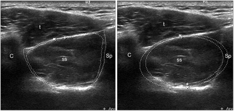
Fig. 2 Calculation of the occupation ratio by sonography reproducing MRI Y-view. C: clavicle; Sp: Scapular spine; t: trapezius; ss: supraspinatus.
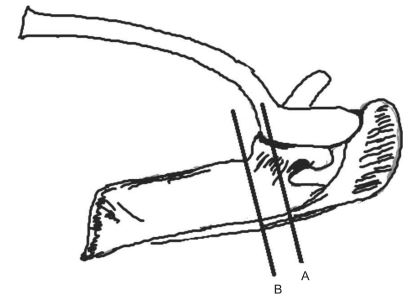
Fig. 3 (A) The sagittal oblique line on the MRI Y-view is surrounded mostly by bony boundaries. (B) The application line of the probe on ultrasonography can be displaced to avoid bony interference.
Cited by 1 articles
-
Reliability of the Supraspinatus Muscle Thickness Measurement by Ultrasonography
Tae Im Yi, In Soo Han, Joo Sup Kim, Ju Ryeon Jin, Jea Shin Han
Ann Rehabil Med. 2012;36(4):488-495. doi: 10.5535/arm.2012.36.4.488.
Reference
-
1. Sheffler LR, Chae J. Neuromuscular electrical stimulation in neurorehabilitation. Muscle Nerve. 2007; 35:562–590. PMID: 17299744.
Article2. Thomazeau H, Boukobza E, Morcet N, Chaperon J, Langlais F. Prediction of rotator cuff repair results by magnetic resonance imaging. Clin Orthop Relat Res. 1997; 344:275–283. PMID: 9372778.
Article3. Lehtinen JT, Tingart MJ, Apreleva M, Zurakowski D, Palmer W, Warner JJ. Practical assessment of rotator cuff muscle volumes using shoulder of rotator cuff msucle volumes using shoulder MRI. Acta Orthop Scand. 2003; 74:722–729. PMID: 14763706.4. Khoury V, Cardinal E, Brassard P. Atrophy and fatty infiltration of the supraspinatus muscle: sonography versus MRI. AJR Am J Roentgenol. 2008; 190:1105–1111. PMID: 18356462.
Article5. Thomazeau H, Rolland Y, Lucas C, Duval JM, Langlais F. Atrophy of the supraspinatus belly, Assessment by MRI in 55 patients with rotator cuff pathology. Acta Orthop Scand. 1996; 67:264–268. PMID: 8686465.
Article6. Gottdiener JS, Livengood SV, Meyer PS, Chase GA. Should echocardiography be performed to assess effects of antihypertensive therapy? Test-retest reliability of echocardiography for measurement of left ventricular mass and function. J Am Coll Cardiol. 1995; 25:424–430. PMID: 7829797.
Article7. Esformes JI, Narici MV, Maganaris CN. Measurement of human muscle volume using ultrasonography. Eur J Appl Physiol. 2002; 87:90–92. PMID: 12012082.
Article8. Reeves ND, Maganaris CN, Narici MV. Ultrasonographic assessment of human skeletal muscle size. Eur J Appl Physiol. 2004; 91:116–118. PMID: 14639480.
Article9. Juul-Kristensen B, Bojsen-Moller F, Holst E, Ekdahl C. Comparison of muscle sizes and moment arms of two rotator cuff muscles measured by ultrsonography and magnetic resonance imaging. Eur J Ultrasound. 2000; 11:161–173. PMID: 10874191.
- Full Text Links
-
- Actions
-
Cited
- CITED
-
- Close
- Share
-
- Similar articles
-
- Reliability of the Supraspinatus Muscle Thickness Measurement by Ultrasonography
- Comparisons of Test-Retest Reliability of Strength Measurement of Gluteus Medius Strength between Break and Make Test in Subjects with Pelvic Drop
- Testing the Reliability of the Pain Color Circle Measurement Tool
- Measurement of Loudness Discomfort Levels as a Test for Hyperacusis: Test-Retest Reliability and Its Clinical Value
- A Psychometric Evaluation of a Korean Version of the Quality of Life in Late-Stage Dementia Scale
