A Case of Primary Choriocarcinoma of the Lung
- Affiliations
-
- 1Division of Pulmonary and Critical Care Medicine, Department of Internal Medicine and Lung Institute, Seoul National University College of Medicine, Seoul, Republic of Korea. ywkim@snu.ac.kr
- KMID: 1970244
- DOI: http://doi.org/10.4046/trd.2006.61.6.578
Abstract
- A primary choriocarcinoma of the lung is extremely rare, and difficult to distinguish from a metastatic choriocarcinoma considering that the lung is also one of the most frequent sites of metastasis. We report a 28-year-old woman patient who was initially misdiagnosed with an ectopic pregnancy and was operated on under the impression of an unidentified malignancy of the lung, which was finally proven to be a choriocarcinoma of the lung. A pelvic examination by a gynecologist, pelvic magnetic resonance imaging and whole body fluorodeoxyglucose positron emission tomography-computed tomography was performed in order to rule out a metastatic choriocarcinoma of the lung. After a curative operation, her serum beta-human chorionic gonadotropin (HCG) level, which was highly elevated in the initial evaluation, had decreased dramatically to the normal range. She is currently being followed up regularly without any evidence of recurrence or elevation of her beta-HCG level.
MeSH Terms
Figure
-

Figure 1 Chest radiograph shows an oval shaped-mass in right hilar area(A). Whole body F-18 FDG Fusion PET-CT shows hypermetabolic lesion in right lower lobe area. The maxSUV of lesion is 6.3(B).

Figure 2 Chest CT and PET-CT shows a lobulating mass with calcification in the right lower lobe superior segment(A: non-contrast CT, B: contrast-CT, C: lung window setting, D: PET-CT).
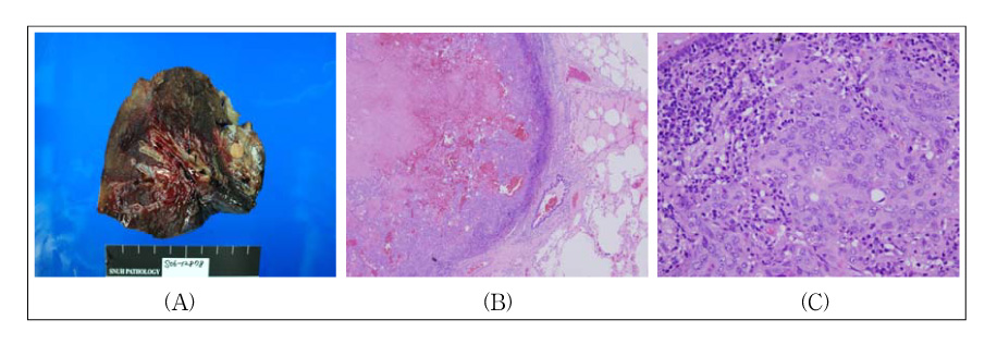
Figure 3 Gross pathologic image of right lower lobectomy specimen is demonstrating a large intraparenchymal choriocarcinoma(A). Necroses and hemorrhages are showed in the central area of tumor components(B; H&E stain, ×40). Tumor nests are composed of the dimorphic population of malignant cytotrophoblasts and syncytiotrophoblasts(C; H&E stain, ×400).
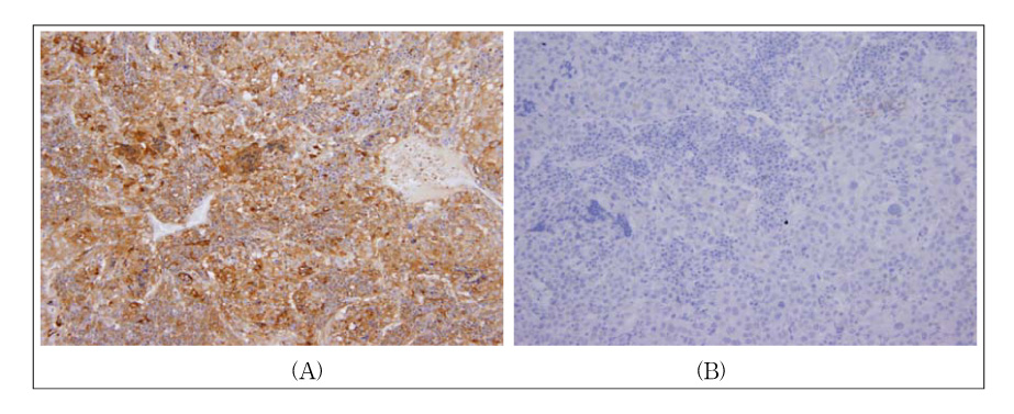
Figure 4 The immunohistochemical staining of the tumor shows positive for β-HCG(A; Immunohistochemical stain, ×200) and negative for TTF-1(B; Immunohistochemical stain, ×200)
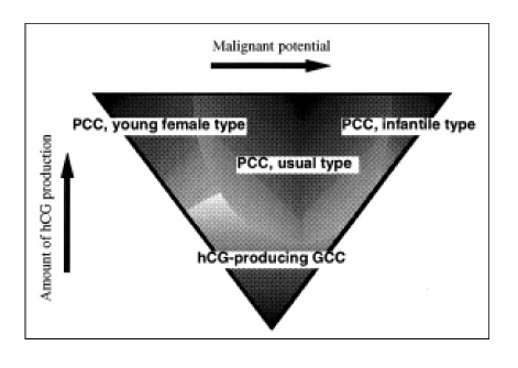
Figure 5 Ikura's5 hypothetical concept on the conformation of the group of primary pulmonary choriocarcinooma (PCC).
Reference
-
1. Umemori Y, Hiraki A, Aoe K, Murakami T, Maeda T, Matsuda E, et al. Primary choriocarcinoma of the lung. Anticancer Res. 2004. 24:1905–1910.2. Yamamoto S, Tanaka H, Takeo H, Yasuda K, Mastukuma S. Primary pulmonary choriocarcinoma combined with adenocarcinoma. Pathol Int. 2006. 56:402–407.3. Pushchak MJ, Farhi DC. Primary choriocarcinoma of the lung. Arch Pathol Lab Med. 1987. 111:477–479.4. Tanimura A, Natsuyama H, Kawano M, Tanimura Y, Tanaka T, Kitazon M. Primary choriocarcinoma of the lung. Human Pathol. 1985. 16:1281–1284.5. Ikura Y, Inoue T, Tsukuda H, Yamamoto T, Ueda M, Kobayashi Y. Primary choriocarcinoma and human chorionic gonadotrophin-producing giant cell carcinoma of the lung: are they independent entities? Histopathology. 2000. 36:17–25.6. Chen HX. Expanding the clinical development of bevacizumab. Oncologist. 2004. 9:Suppl 1. 27–35.7. Toda S, Inoue Y, Ishino T, Yonemitsu N, Terayama K, Miyabara S, et al. A rare case of primary pulmonary choriocarcinoma in a male: immunohistochemical detection for human chorionic gonadotropin, epidermal growth factor (EGF) and EGF-receptor. Endocr J. 1995. 42:655–659.8. Shintaku M, Hwang MH, Amitani R. Primary choriocarcinoma of the lung manifesting as diffuse alveolar hemorrhage. Arch Pathol Lab Med. 2006. 130:540–543.9. Fusco FD, Rosen SW. Gonadotropin-producing anaplastic large-cell carcinomas of the lung. N Engl J Med. 1966. 275:507–515.10. Sagaster P, Zojer N, Dekan G, Ludwig H. A paraneoplastic syndrome mimicking extrauterine pregnancy. Ann Oncol. 2002. 13:170–172.11. Tsai JR, Chong IW, Hung JY, Tsai KB. Use of urine pregnancy test for rapid diagnosis of primary pulmonary choriocarcinoma in a man. Chest. 2002. 121:996–998.12. Aparicio J, Oltra A, Martinez-Moragon E, Llorca C, Gomez-Aldaravi L, Paster M. Extragonadal nongestational choriocarcinoma involving the lung: a report of three cases. Respiration. 1996. 63:251–253.13. Sridhar KS, Saldana MJ, Thurer RJ, Beattie EJ. Primary choriocarcinoma of the lung: report of a case treated with intensive multimodality therapy and review of the literature. J Surg Oncol. 1989. 41:93–97.14. Numnum TM, Leath CA 3rd, Straughn JM Jr, Conner MG, Barnes MN 3rd. Occult choriocarcinoma discovered by positron emission tomography/computed tomography imaging following a successful pregnancy. Gynecol Oncol. 2005. 97:713–715.15. van Nostrand KM, Lucci JA 3rd, Liao SY, di Saia PJ. Primary lung choriocarcinoma masquerading as a metastatic gestational neoplasm. Gynecol Oncol. 1994. 53:361–365.
