Clin Endosc.
2015 Jul;48(4):332-335. 10.5946/ce.2015.48.4.332.
Simultaneous Esophageal and Gastric Metastases from Lung Cancer
- Affiliations
-
- 1Department of Internal Medicine, Seoul National University Hospital, Seoul National University College of Medicine, Seoul, Korea. jpim0911@snu.ac.kr
- KMID: 1964278
- DOI: http://doi.org/10.5946/ce.2015.48.4.332
Abstract
- We report of a patient with metastatic adenocarcinoma of the esophagus and stomach from lung cancer. The patient was a 68-year-old man receiving radiotherapy and chemotherapy for stage IV lung cancer, without metastases to the gastrointestinal (GI) tract at the time of the initial diagnosis. During the treatment period, dysphagia and melena newly developed. Upper GI endoscopy revealed geographic erosion at the distal esophagus and multiple volcano-shaped ulcers on the stomach body. Endoscopic biopsy was performed for each lesion. To determine whether the lesions were primary esophageal and gastric cancer masses or metastases from the lung cancer, histopathological testing including immunohistochemical staining was performed, and metastasis from lung cancer was confirmed. The disease progressed despite chemotherapy, and the patient died 5 months after the diagnosis of lung cancer. This is a case report of metastatic adenocarcinoma in the esophagus and stomach, which are very rare sites of spread for lung cancer.
MeSH Terms
Figure
-
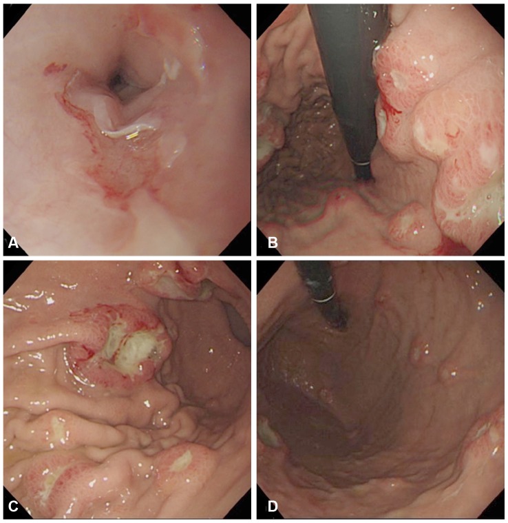
Fig. 1 Endoscopic findings showing gastrointestinal metastases. (A) Geographic erosion is observed at the distal esophagus 35 cm from the upper incisors, with epithelial break and mild hemorrhagic change. There is no evidence of active bleeding. (B-D) The stomach shows numerous volcano-shaped sessile masses with central umbilication, which vary in size.
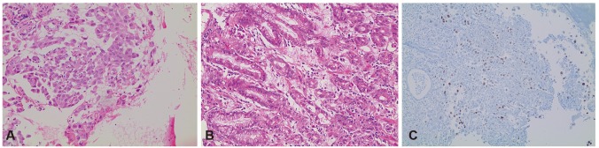
Fig. 2 Histological and immunohistochemical staining results. The histologic results of the esophageal and gastric metastatic lesions (A, H&E stain, ×200) and right lower paratracheal lymph node (4R) lesions (B, H&E stain, ×100) all showed adenocarcinomas. The immunohistochemical staining of the stomach lesions was positive for thyroid transcriptional factor-1 (C, ×100).
Reference
-
1. Hsu PK, Shai SE, Wang J, Hsu CP. Esophageal metastasis from occult lung cancer. J Chin Med Assoc. 2010; 73:327–330. PMID: 20603092.
Article2. Antler AS, Ough Y, Pitchumoni CS, Davidian M, Thelmo W. Gastrointestinal metastases from malignant tumors of the lung. Cancer. 1982; 49:170–172. PMID: 6274500.
Article3. Jemal A, Bray F, Center MM, Ferlay J, Ward E, Forman D. Global cancer statistics. CA Cancer J Clin. 2011; 61:69–90. PMID: 21296855.
Article4. Hung TI, Chu KE, Chou YH, Yang KC. Gastric metastasis of lung cancer mimicking an adrenal tumor. Case Rep Gastroenterol. 2014; 8:77–81. PMID: 24748862.
Article5. Kobayashi O, Murakami H, Yoshida T, et al. Clinical diagnosis of metastatic gastric tumors: clinicopathologic findings and prognosis of nine patients in a single cancer center. World J Surg. 2004; 28:548–551. PMID: 15366743.
Article6. McNeill PM, Wagman LD, Neifeld JP. Small bowel metastases from primary carcinoma of the lung. Cancer. 1987; 59:1486–1489. PMID: 3028602.
Article7. Yoshimoto A, Kasahara K, Kawashima A. Gastrointestinal metastases from primary lung cancer. Eur J Cancer. 2006; 42:3157–3160. PMID: 17079136.
Article8. Sileri P, D'Ugo S, Del Vecchio Blanco G, et al. Solitary metachronous gastric metastasis from pulmonary adenocarcinoma: report of a case. Int J Surg Case Rep. 2012; 3:385–388. PMID: 22634567.
Article9. Kadakia SC, Parker A, Canales L. Metastatic tumors to the upper gastrointestinal tract: endoscopic experience. Am J Gastroenterol. 1992; 87:1418–1423. PMID: 1415098.10. Pomerantz H, Margolin HN. Metastases to the gastrointestinal tract from malignant melanoma. Am J Roentgenol Radium Ther Nucl Med. 1962; 88:712–717.11. Scobie BA. Malignant gastric ulcer due to metastasis. Australas Radiol. 1966; 10:119–123. PMID: 5939878.
Article12. Hsu CC, Chen JJ, Changchien CS. Endoscopic features of metastatic tumors in the upper gastrointestinal tract. Endoscopy. 1996; 28:249–253. PMID: 8739742.
Article13. Oda , Kondo H, Yamao T, et al. Metastatic tumors to the stomach: analysis of 54 patients diagnosed at endoscopy and 347 autopsy cases. Endoscopy. 2001; 33:507–510. PMID: 11437044.
Article14. Reis-Filho JS, Carrilho C, Valenti C, et al. Is TTF1 a good immunohistochemical marker to distinguish primary from metastatic lung adenocarcinomas? Pathol Res Pract. 2000; 196:835–840. PMID: 11156325.
Article15. Yang CJ, Hwang JJ, Kang WY, et al. Gastro-intestinal metastasis of primary lung carcinoma: clinical presentations and outcome. Lung Cancer. 2006; 54:319–323. PMID: 17010474.
Article16. Rossi G, Marchioni A, Romagnani E, et al. Primary lung cancer presenting with gastrointestinal tract involvement: clinicopathologic and immunohistochemical features in a series of 18 consecutive cases. J Thorac Oncol. 2007; 2:115–120. PMID: 17410025.
Article
- Full Text Links
-
- Actions
-
Cited
- CITED
-
- Close
- Share
-
- Similar articles
-
- A Case of Esophageal Adenocarcinoma Metastasized to the Scalp
- Simultaneous Laparoscopy-Assisted Resection for Colorectal Cancer and Metastases
- Resection for Pancreatic Cancer Lung Metastases
- Cutaneous Metastasis from Prostatic Cancer
- A Case of Cavitary Pulmonary Metastases of Primary Cavitary Lung Cancer
