Tumefactive Immunoglobulin G4-Related Disease Involving the Dura Mater: A Case Report and Literature Review
- Affiliations
-
- 1Department of Radiology, Hanyang University Hospital, Seoul, Korea. jyjy133@naver.com
- 2Department of Radiology, Hanyang University Guri Hospital, Guri, Korea.
- 3Department of Neurosurgery, Hanyang University Hospital, Seoul, Korea.
- 4Department of Pathology, Hanyang University Hospital, Seoul, Korea.
- KMID: 1964261
- DOI: http://doi.org/10.3348/jksr.2015.73.2.108
Abstract
- Immunoglobulin G4 (IgG4)-related disease is a well-known disorder characterized by an inflammatory reaction with an increase in the number of IgG4-positive plasma cells associated with sclerosis. IgG4-related disease often affects the dura mater with a pattern of diffuse thickening when the central nervous system is involved. However, some nodular dural thickening requires discrimination from tumors because of obviously different treatment options. We report of a case of IgG4-related disease with tumefactive dural involvement.
MeSH Terms
Figure
-
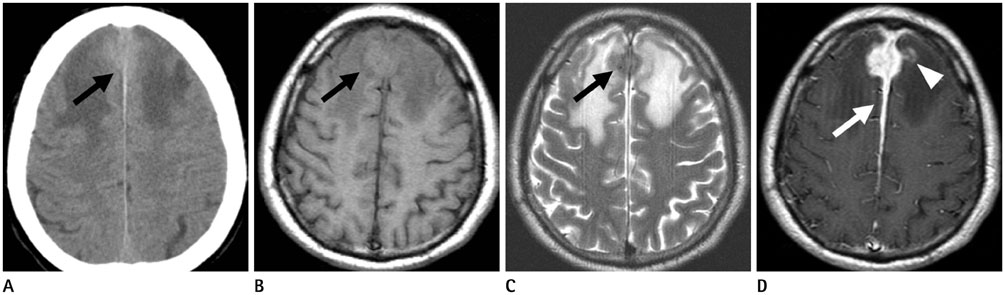
Fig. 1 Computed tomography (CT) and magnetic resonance imaging of immunoglobulin G4-related disease with tumefactive dural involvement in a 57-year-old man. Initial non-enhanced CT (A) demonstrates a slightly hyperdense mass (black arrow) along the anterior falx. The mass (black arrow) shows intermediate signal intensity on T1- (B) and low signal intensity on T2-weighted images (C) with extensive perilesional parenchymal edema. A post-contrast T1-weighted image (D) shows, diffuse thickening and enhancement of dura mater (white arrow) at the anterior falx cerebri and parasagittal region. The mass shows homogeneous enhancement with a jagged border and co-involvement of adjacent brain parenchyma (white arrowhead).
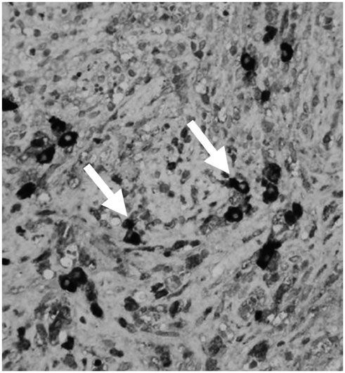
Fig. 2 Immunoglobulin G4 (IgG4) immunohistochemical staining (× 400) shows infiltration of abundant IgG4-positive plasma cells (white arrows).
Cited by 1 articles
-
A Case of IgG4 Related Pachymeningitis
Ji In Kim, Jin Taek Song, Hyeong Ju Kwon, Ji-Yong Lee
J Neurocrit Care. 2016;9(2):162-165. doi: 10.18700/jnc.160075.
Reference
-
1. Moss HE, Mejico LJ, de la Roza G, Coyne TM, Galetta SL, Liu GT. IgG4-related inflammatory pseudotumor of the central nervous system responsive to mycophenolate mofetil. J Neurol Sci. 2012; 318:31–35.2. Lindstrom KM, Cousar JB, Lopes MB. IgG4-related meningeal disease: clinico-pathological features and proposal for diagnostic criteria. Acta Neuropathol. 2010; 120:765–776.3. Lee LK, Sahani DV. Autoimmune pancreatitis in the context of IgG4-related disease: review of imaging findings. World J Gastroenterol. 2014; 20:15177–15189.4. Wallace ZS, Carruthers MN, Khosroshahi A, Carruthers R, Shinagare S, Stemmer-Rachamimov A, et al. IgG4-related disease and hypertrophic pachymeningitis. Medicine (Baltimore). 2013; 92:206–216.5. Takeuchi S, Osada H, Seno S, Nawashiro H. IgG4-Related Intracranial Hypertrophic Pachymeningitis: a case report and review of the literature. J Korean Neurosurg Soc. 2014; 55:300–302.6. Lee YC, Chueng YC, Hsu SW, Lui CC. Idiopathic hypertrophic cranial pachymeningitis: case report with 7 years of imaging follow-up. AJNR Am J Neuroradiol. 2003; 24:119–123.7. D'Andrea G, Trillò G, Celli P, Roperto R, Crispo F, Ferrante L. Idiopathic intracranial hypertrophic pachymeningitis: two case reports and review of the literature. Neurosurg Rev. 2004; 27:199–204.8. Lu LX, Della-Torre E, Stone JH, Clark SW. IgG4-related hypertrophic pachymeningitis: clinical features, diagnostic criteria, and treatment. JAMA Neurol. 2014; 71:785–793.9. Hamid HA, Gee KY, Muhammad R, Abd Rahman ZA, Das S. Dural metastasis mimicking meningioma: an interesting case. Acta Medica (Hradec Kralove). 2009; 52:19–22.10. Smirniotopoulos JG, Murphy FM, Rushing EJ, Rees JH, Schroeder JW. Patterns of contrast enhancement in the brain and meninges. Radiographics. 2007; 27:525–551.
- Full Text Links
-
- Actions
-
Cited
- CITED
-
- Close
- Share
-
- Similar articles
-
- Unusual Manifestation of Immunoglobulin G4-Related Disease Involving the Upper Arm: A Case Report
- Immunoglobulin G4-Related Hypertrophic Pachymeningitis with Skull Involvement
- Immunoglobulin G4-Related Disease Involving Various Head and Neck Regions: A Case Report
- Erdheim–Chester Disease Involving the Biliary System and Mimicking Immunoglobulin G4-Related Disease: A Case Report
- IgG4-Related Intracranial Hypertrophic Pachymeningitis : A Case Report and Review of the Literature
