Delayed Dural Arteriovenous Fistula after Microvascular Decompression for Hemifacial Spasm
- Affiliations
-
- 1Department of Neurosurgery, Yonsei University College of Medicine, Seoul, Korea. jchang@yuhs.ac
- KMID: 1956524
- DOI: http://doi.org/10.3340/jkns.2014.56.2.168
Abstract
- Dural arteriovenous fistula (AVF) is very rare, acquired lesion that may present with intracranial hemorrhage or neurological deficits. The etiology is not completely understood but dural AVF often has been associated with thrombosis of the involved dural sinuses. To our knowledge, this is the first well documented intracranial hemorrhage case caused by dural AVF following microvascular decompression for hemifacial spasm. A 49-year-old male patient had left microvascular decompression of anterior inferior cerebellar artery via retrosigmoid suboccipital craniotomy. The patient was in good condition without any residual spasm or surgery-related complications. However, after 10 months, he suffered sudden onset of amnesia and dysarthria. Computed tomography and magnetic resonance imaging revealed the presence of dural AVF around the left transverse-sigmoid sinus. The dural AVF was treated with Onyx(R) (ev3) embolization. At the one-year follow up visit, there were no evidence of recurrence and morbidity related to dural AVF and its treatment. This case confirms that the acquired etiology of dural AVF may be associated with retrosigmoid suboccipital craniotomy for hemifacial spasm, even though it is an extremely consequence of this procedure.
Keyword
MeSH Terms
Figure
-
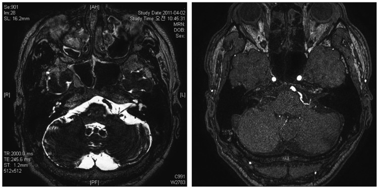
Fig. 1 Preoperative magnetic resonance image show that left anterior inferior cerebellar artery compressed left facial nerve at the root exit zone.
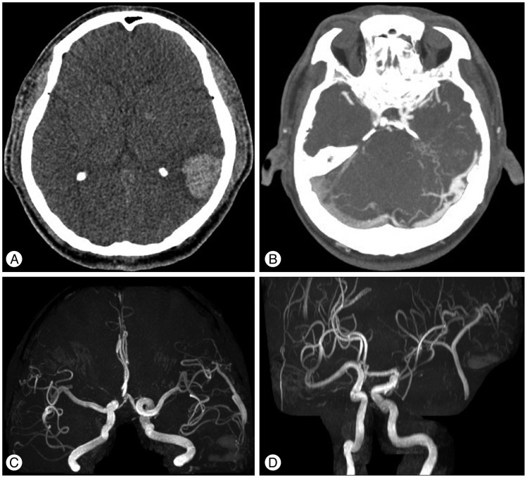
Fig. 2 A : Computed tomography (CT) shows the high density lesion on the left temporoparietal subcortical area. B : CT angiography shows the abnormal enhanced vessels on left transverse-sigmoid sinus. C and D : MRAs show dural arteriovenous fistula that has occipital artery as a feeder and draining transverse-sigmoid sinus.
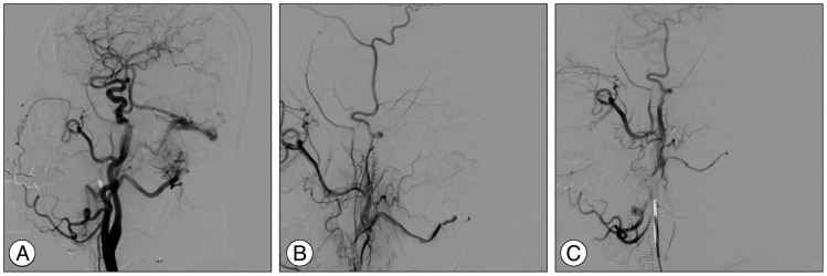
Fig. 3 A : Pre-embolizational left external carotid artery angiographic image shows the dural AVF with feeders from left occipital artery. B : Post-embolizational angiographic image shows nearly complete occlusion of left sigmoid sinus dural arteriovenous fistula (AVF). C : There is no residual dural AVF at the follow-up angiography obtained 12 months after embolization.
Reference
-
1. Bink A, Berkefeld J, Kraus L, Senft C, Ziemann U, du Mesnil de Rochemont R. Long-term outcome in patients treated for benign dural arteriovenous fistulas of the posterior fossa. Neuroradiology. 2011; 53:493–500. PMID: 20737143.
Article2. Brown RD Jr, Wiebers DO, Nichols DA. Intracranial dural arteriovenous fistulae : angiographic predictors of intracranial hemorrhage and clinical outcome in nonsurgical patients. J Neurosurg. 1994; 81:531–538. PMID: 7931586.
Article3. Caragine LP, Halbach VV, Dowd CF, Ng PP, Higashida RT. Parallel venous channel as the recipient pouch in transverse/sigmoid sinus dural fistulae. Neurosurgery. 2003; 53:1261–1266. discussion 1266-1267. PMID: 14633292.
Article4. Chaudhary MY, Sachdev VP, Cho SH, Weitzner I Jr, Puljic S, Huang YP. Dural arteriovenous malformation of the major venous sinuses : an acquired lesion. AJNR Am J Neuroradiol. 1982; 3:13–19. PMID: 6800236.5. Cognard C, Gobin YP, Pierot L, Bailly AL, Houdart E, Casasco A, et al. Cerebral dural arteriovenous fistulas : clinical and angiographic correlation with a revised classification of venous drainage. Radiology. 1995; 194:671–680. PMID: 7862961.
Article6. Gardner WJ, Sava GA. Hemifacial spasm-a reversible pathophysiologic state. J Neurosurgery. 1962; 19:240–247.
Article7. Hanaoka M, Matsubara S, Satoh K, Nagahiro S. Dural arteriovenous fistulae after cerebral infarction : report of two cases. Neurosurgery. 2011; 68:E575–E579. discussion E580. PMID: 21135717.8. Houser OW, Baker HL Jr, Rhoton AL Jr, Okazaki H. Intracranial dural arteriovenous malformations. Radiology. 1972; 105:55–64. PMID: 5057316.
Article9. Jannetta PJ, Abbasy M, Maroon JC, Ramos FM, Albin MS. Etiology and definitive microsurgical treatment of hemifacial spasm. Operative techniques and results in 47 patients. J Neurosurg. 1977; 47:321–328. PMID: 894338.10. Jiang C, Lv X, Li Y, Wu Z. Transarterial Onyx packing of the transverse-sigmoid sinus for dural arteriovenous fistulas. Eur J Radiol. 2011; 80:767–770. PMID: 21030176.
Article11. Kida Y. Radiosurgery for dural arteriovenous fistula. Prog Neurol Surg. 2009; 22:38–44. PMID: 18948718.
Article12. Kim JP, Chung JC, Chang WS, Chung SS, Chang JW. Outcomes of surgical treatment for hemifacial spasm associated with the vertebral artery : severity of compression, indentation, and color change. Acta Neurochir (Wien). 2012; 154:501–508. PMID: 22160400.
Article13. Kimura T, Shojima M, Koizumi T, Fukaya S, Ichikawa Y, Morita A. Surgical treatment of a dural arteriovenous fistula in the superior petrosal sinus of an elderly patient with minimal suboccipital craniotomy : case report. Neurol Med Chir (Tokyo). 2009; 49:465–467. PMID: 19855143.
Article14. Loumiotis I, Lanzino G, Daniels D, Sheehan J, Link M. Radiosurgery for intracranial dural arteriovenous fistulas (DAVFs) : a review. Neurosurg Rev. 2011; 34:305–315. discussion 315. PMID: 21584687.
Article15. Lv X, Jiang C, Li Y, Liu L, Liu J, Wu Z. The limitations and risks of transarterial Onyx injections in the treatment of grade I and II DAVFs. Eur J Radiol. 2011; 80:e385–e388. PMID: 20837384.
Article16. Lv X, Jiang C, Li Y, Wu Z. Embolization of intracranial dural arteriovenous fistulas with Onyx-18. Eur J Radiol. 2010; 73:664–671. PMID: 19144481.
Article17. Miller LE, Miller VM. Safety and effectiveness of microvascular decompression for treatment of hemifacial spasm : a systematic review. Br J Neurosurg. 2012; 26:438–444. PMID: 22168965.
Article18. Nabors MW, Azzam CJ, Albanna FJ, Gulya AJ, Davis DO, Kobrine AI. Delayed postoperative dural arteriovenous malformations. Report of two cases. J Neurosurg. 1987; 66:768–772. PMID: 3572502.19. Sakaki T, Morimoto T, Nakase H, Kakizaki T, Nagata K. Dural arteriovenous fistula of the posterior fossa developing after surgical occlusion of the sigmoid sinus. Report of five cases. J Neurosurg. 1996; 84:113–118. PMID: 8613817.
Article20. Schaller B. Trigeminocardiac reflex. A clinical phenomenon or a new physiological entity? J Neurol. 2004; 251:658–665. PMID: 15311339.
- Full Text Links
-
- Actions
-
Cited
- CITED
-
- Close
- Share
-
- Similar articles
-
- Dural Arteriovenous Fistula Involving an Isolated Sinus Treated Using Transarterial Onyx Embolization
- Generalized Convulsion and Delayed Emergence following Microvascular Decompression Surgery under General Anesthesia: A case report
- Intraoperative monitoring of microvascular decompression in hemifacial spasm
- Delayed Unilateral Soft Palate Palsy without Vocal Cord Involvement after Microvascular Decompression for Hemifacial Spasm
- Microvascular triple decompression for combined simultaneous trigeminal neuralgia, hemifacial spasm, and pulsatile tinnitus due to separate offending vessels
